Bone, joints and soft tissues part 2 Flashcards
(27 cards)
AKA exostosis
most common benign bone tumor in early adults (85%)
younger in AD multiple hereditary exotoses and men>female
LOF of EXT1 or EXT 2 in sporadic osteochondromas, encode synthrsis of heparin sulfate gylcosaminoglycans
located: metaphysis near growth plate of long bone, especially the knee
painful if impinge a nerve
multiple hereditary exostosis can progress to chondrosarcoma
morphology: mushroom shaped surface protrusion, covered by perichondrium overlying a hyaline cartilage cap
the cap have the appearance of a disorganized growth plate and undergo endochondrial ossification. the stalk cortex and medullary cavities are in continuation with the cortex and marrow cavity of the underlying bone

osteochondroma

benign hyaline cartilage, solitary metaphyseal lesions (hands and feet) in 20-50 yo
may be within medullary cavity (enchondroma)
isocitrate dehydrogenase gene IDH1 and IDH2 mutation
morphology: <3cm, gray blue and translucent
microscopically: nodules of hyaline cartilage, there is pheripheral enchondral ossification and central calcification and necrosis
clinical presentation: asymptomatic but may cause bone deformities (esp. in enchondromatosis) pain, fractures. radiologic image reveal well circumscribed oval lucencies with thin rim of radiodense bone, matrix calcification is detected as irregular opacification
evolvment to sarcoma is frequent in enchondromatosis and patient with Maffucci syndrome are at risk of ovarian cancinoma and CNS gliomas

chondroma/enchondroma
Ollier syndrome-multiple enchrondroma
mafucci syndrome=multiple enchondroma + angiomas–> increase risk of chondrosarcoma and other malignancies
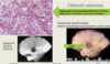
second most common malignant matrix pruducing tumor bone (osteosarcoma first)
M>F
usually in 40s or olders
axial skeleton (pevis, shoulders, ribs)
invade locally, painful enlarging mass, may metastasize
grade 3 spread hematogeously to lungs
radiologic studies shows endosteal scalloping and flocculent-appearing matrix calcification. this is a slow growing tumor
morphology: tumors are lubulated, fray, glistening and semitranslucent necrosis and spotty calcification
microscopically most tumors are low grade and classified as intramedullary (90%) or juxacortical
the grades are based cellularity, cytologic atypia and miotic activity)

chondrosarcoma
painful, usually at worse at night, classically responds to aspirin and NSAIDs
excess PGE2 by osteoblasts in young men in teens and 20s
location: appendicular skeletal = 50% in femus or tibia
<2 cm in greatest dimension (>2cm = osteoblastoma)
have thick rind of reactive cotical bone= radiographic clue
gross: round-oval masses of hemorrhagic, gritty, tan tissue
microscopically: central nidus of translucent woven bone (nidus) surrounded by rim of osteoblasts
tx by radiofrequency ablation
Osteoid osteoma
bigger than osteoid osteoma >2cm
involves the posterior spine
no bony reaction
achy pain that does not respond to aspirin
Tx: curetted or excised en bloc
malignancy is rare
ostoblastoma
malignant mesenchymal cells produce bone matrix (most common primary malignant tumor)
present as painful enlarging mass or pathologic fracture
bimodal 75 <20yo males 75% around the knee, 2nd peak is in older males with pagets, prior radiation (secondary)
RB gene mutation increase risk and seen in 70% of sporadic cases
TP53 (Li-Graumani syndrome) mutation
INK4a gene inactivated
MDM2 adn CDK4 inhibit p53 and Rb, respectively in Low Grade osteosarcomas
xray- mixed lytic and blastic mass
-codman triangle (elevation of peristeum)
spread early into lungs (hematogenously) can spread to bones or brain who die with tumor
morphology: large destructive, tan-white, gritty, sometime bloody and cystic mass
microscopically: frrequently have coarse, lace-like pattern
clinical course arise in medullary cavity of metaphyses of long bone (1/2 occur around knee)

osteosarcoma

80% are > 20yo, striking predilection for whites
second most common sarcoma in children
ESFT is undifferentiated with neural differentiation, primitive neuroectodermal tumor (PNET)
locations: diaphyses of long bones esp femur and flat bones
arises in medullary cavity, invades cortex
pathogenesis: t(11;22)(q24:q12) translocation fusing EWS gene to the FL1 gene
morphology: invade cortex and penetrate the periosteum to produce a tanowhite soft tissue mass with focal hemorrhage and necrosis
microscopically: small blue round cell tumor, homer-Wright pseudorosettes (tumor cells arrayed in a circle about a central fibrillary space) -indicate neural differentiation
clinical course: painful enlarging mass, frequently tender, warm and swollen (fever, increase ESR)-mimic infection due leukocytosis, periosteal rxn–> reactive bone in onion skin fashion on xray

Ewing Sarcoma Family Tumor (ESFT)

benign proliferation of fibrous tissue and bone that do not mature (developmental arrest)
70% monostotic (usually incidental finding due to pain, fracture or discrepencies in limb length, craniofacial), 27% polyostotic (crippling deformities- craniofacial shoulder, pelvic gridle are common)
mostly early adolescence (stops enlarging as growth plate closes)
M=F
1/3 craniofacial bones, 1/3 femur or tibia, and 1/3 ribs
ground glass appearance, well defined margins radiographically
- what is McCune-Albright disease?
- what is Mazabraud syndrome?
fibrous dysplasia
- phenotype depend on 1) when in development the mutation is acquired. 2) fate of the cell harboring the mutation (if osteoblast precursor mutation during skeletal formation then that would result in monostotic fibrous dysplasia, whereas mutation during embrypgenesis results in McCune-Albright)
1. mutation during emryogenesis, bone lesions, unilateral, pigmented skin lesion (cafe au lait) same side, with endocrine disorders - precocious puberty in females (& other endocrinopathies- hyperthyroidism , GH-secreting pituitary adenoma, primary adrenal hyperplasia)
GNAS mutation-GNAS gene makes the alpha G subunit of the GPCR-help stimulte adenylate cyclase
this is known to to help regulate glands like thyroid, pituitary, ovaries, testes and adrenal. can play role to help regulate the development of bone
-
fibrous dysplasia (usually polyostotic) multiple skeletal deformities in childhood
- soft tissue myxomas (intramuscular) in adulthood in same anatomic sites

osteoclastoma multinucleated ostoclase-type giant cells-must consider brown tumor of hyperparathyroidism
20-40, benign but locally aggressive
derived from monocyte-macrophage derivation
arise in epiphyses, may extend into metaphysis (knee: distal femur, proximal tibia–> arthritis0like symptoms)
morphology: tumor are large and red-brown with infrequent cystic degeneration
microscopically: plump, uniform mononuclear cell constitute that proliferating component of the tumor in a background of ostoclast-like multinucleated giant cells

giant cell tumor of bones
most common form of skeletal maligancy
adult: 75% from prostate, breast, kidney, lungs
kids: neuroblastoma, Wilms, osteosarcoma, Ewing, rhabdomyosarcoma
multifocal except kidney and thyroid usually solitary
axial skeleton: red marrow facilitates implantation and growth
small bones of the hands and feet=lung, kidney, colon
radiographic= lytic- kidney, lung, GI, melanoma
blastic: prostatic adenocarcinoma (ostosclerosis)
metastatic tumors
primary or idiopathic= aging, oligoarticular, exponentially increased >50
hands and knees women, hips in men
secondary osteoarthritis- predisposing condition: jt deformity, trauma, obesity, DM, hemocromatosis, thyroid, acromaly Charcot
dz of cartilage due to wear and tear
evening stiffness, crepitus, limited range of motion, gets worse with use
morphology: dislodged fragment of cartilages and subchondral bone formation (joint mice), subchondrondal bone exposed and rubbed smooth= eburnation (bone on bone pain), bony overgrowth (osteophytes) capped by cartilage also develop at egde of articular cartilage; Herberden nodes are osteophytes of the distal interphalangeal joints (common in women)
the osteophytes can compress nerve roots and cause radicular pain, neuro deficits
microscopically: water content of matrix increase and concentration of proteoglycan decrease–> crack in matrix

osteoarthritis/degenerative joint disease

peaks 2nd-4th decades affecting more women due to autoimmune, nonsuppurative proliferative and inflammatory synovitis
destruction of articular cartilage
- ankylosis: stiffness of joint due to abnormal adhesion and rigidity of the bones of the joint, which may be a result of injury or disease
symmetrical distribution, small joints
- swollen, warm, painful
morning stiffness or gelling after rest lasting >1hr in active dz
Signs
Boutonniere: deformity of finger: hyperextension of DIP, flexion of PIP
Swan-neck- hyperextension of PIP, flexion of DIP
ulnar deviation fingers: decrease jt space, periarticular bony erosion
radial deviation of the waist
RA pathology
pannus: edematous, thickened, hyperplastic synovium-can bridge apposing bones to form a fivrous ankylosis that will eventually ossify
synovial hypertrophy with villi
lumphoid aggregates
Rheumatoid nodule: extensor surfaces at pressure points (firm nontender nodules)
Citrullinated peptides include citrullinated fibrogen, type 2 collagen, alpha-anolase, vimentin. antiboies toward these peptide can act as RA markers, elevated anti-CCP contribute to chronicity
pts with high titer of rheumatoid factor are at increased risk of small-medium sized vessel vasculitis
serum immunoglobulin IgM or IgA that binds to Fc region are called rheumatoid factors

Reumatoid arthritis (RA)

autoimmune, T-cell response initiated by environmental factors
pathologic changes in ligamentous attachments, rather than synovium
- negative RF associated with HLA-B27
ankylosing spndyliti (HLA-B27)- vertebrae and acroiliac joints 20-30m uveitis, aortitis, amykoidosis are complications progressive, 1/3 of pts have peripheral jt involvement (hips, knees, shoulders)
reactive arthritis-1) arthritis, 2) urethritis or cervicitis, 3) conjunctivties (triad)
- 80% HLA-B27, MEN 20-30, also indv with HIV, prior GI (shigella, salmonella, Yersinia, Campylobacter) or GU (chlamydia) infection, sx several weeks later
ankles, knees, feet, in asymmetric pattern, severe chronic spine involvement
extraarticular: conjuntivitis, cardiac conduction abn, aortic regurg.

seronegative spondyloarthropathies
Enteritis associated arthritis: Yersiia, Salmonella, Shiella, Campylobacter
- LPS
psoriatic arthritis: peripheral (hand and feet) and axial hts, ligaments and tendons
- HLA-B27 and HLA-Cw6
30-50 10% of psoriatic population
pencil in cup deformity,, DIP, asymmetrical in 50%
psoriasis: nails can turn thick, roughm and rigid. theu discolor or develop pits. in some cases, the nail can separate fromt he nail bed (known as onycholysis)
microorganism seed joints via hematogenous dissemination
direct inoculation or contiguous spread soft tissue abscess or focus of osteomyelitis
rapid joint destruction–> permanent deformities
infectious arthritis
<2yo H. influenza arthritis
older kids have staph areus, Gonococcus late adolescent, early adult
- gonococcus is more prevalent in sexually active women
- MAC deficiency (C6-7) = susceptible
acutely painful, swollen ht, restricted range of motion-fever leukocytosis and increase ESR
90% non-gonococcal cases involve single joint
- Knew> hips> shoulders> elbow> wrist> sternoclavicular jts
jt aspiration diagnostic, purulent fluid
mycobacterial: insidious onset, gradual progressive pain (hip> knee> ankle)
lyme disease: Borrelia burgdorferi, 60-80% develop days or weeks after skin lesion (knee> shoulders> elbows > ankles)
viral: HIV, HBV, HCV, EBV, parvovirus B19, rubella
suppurative arthritis
transient attacks of acute arthritis initiated by crystallization of monosodium urate (MSU) within the joint
overproduction or reduced excretion (plasma levels >6.8mg/dl)
other factors in addition ro hyperuricemia lead to symptomatic gout:
- age (after 20-30 years of hyperuricemia)
- genetic
- heavy alcohol consumptions
- obesity
- drugs *thiazide diuretics (decrease urate excretion)
lead toxicity (saturnine gout)

gout


aka chondrocalcinosis
>50 yo, increases with increasing age
secondary: associated with previous joint damage, hyperparathyriodism, hemochromatosis, hypothyroidism, diabetes, ochronosis
crystals form chalky, white friable deposits
histoL oval blue-purple aggregates
individual crystals are rhomboid and are positively birefringent
calcium pyrophosphate crystal deposition (CPPD) aka pseudo gout
- joint capsule or tendon sheath 1.-1,5 cm cyst
location: wrise, firm fluctuant pea-sized translucent nodule
result of cystic or myxoid degeneration, cyst wall lacks cell lining
what is this?
herinationof synovium through joint capsule
what is this?
what if this cyst appear in popliteal space, rheumatoid arthritis
what is this?
- ganglion cyst
- synovial cyst
- Baker cyst
benign tumor of adipose tissue (soft, mobile, painless)
most common soft tissue tumor of adults
extremities and trunk
histology: encapsulated mass of normal appearing adipose tissue

lipoma
common malignant ST tumor of adult 50-60yo
deep soft tissue of proximal extremities and retroperitoneum
12q13-q15 and t(12;16)- one key gene in the amplified region of chromosome is MDM2
most common is the myxoid (intermediate) histologic variant

fibrous tumors
self-limited fibroblastic and myofibroblastic proliferation in young adults upper extremity
t(17;22)–> MYH9-USP6 fusion gene = clonal, self limited, proliferation
spontaneous regresses
what is this?
infiltrative, local deformity M>F
palmar (Dupuyren contrature: slow;y progressive flexion contrature of 4-5th fingers
plantar-young adult, unilateral, no contractures
penile: (peyronie disease) dorsolateral aspect of penis may –> abnormal curvature (painful)
what is this?
Desmoid tumor-large, infiltrative painful- in teen-30s most women
mutation of APC or B-catenin genes–> increase Wnt signaling
familial adenomatoous polyposis (Gardner syndrome) who have germline APC mutation are predisposed this condition. what is it?
- nodular fasciitis
- fibromatoses
- deep fibromatosis

adults-deep-seated slow growing mass in thigh or trunk of young or middle aged adults
infantile <2 yo
often congenital
arise in extremitiesl smaller, more superficial
strikingly uniform, spindle cells arranged in herringbone fascicles

fibrosarcoma


