Bone histology Flashcards

a – Volkmann canals
b – perforating/Sharpey fibres
c – periosteum
d – outer circumferential lamella
e – osteocytes
f – intermediate lamellae
g – osteons
h – inner circumferential lamella
i – endosteum
j – bone marrow
k – spongey substance/substantia spongiosa
l – compact substance/substantia compacta

a – osteocytes
b – lamellae of osteons
c – endosteum
d – inner circumferential lamella
e – bone marrow
f – Haversian vessels
g – periosteum
h – perforating/Sharpey fibres

a – bone marrow
b – endosteum
c – inner circumferential lamella
d – Haversian vessels
e – lamellae of osteons
f – osteocytes
g – periosteum
h – perforating/Sharpey fibres
i – Volkmann canals
a – Haversian vessels
b – lamellae of osteons
c – endosteum
d – Volkmann canals
e – bone marrow
What is bone?
A matrix of intercellular material surrounding widely separated cells
What is bone comprised of?
– 50% crystallised mineral
– Hydroxyapatite (Ca phosphate and Ca hydroxide) Ca10(PO4)6(OH)2
– Magnesium hydroxide
– Fluoride
– Sulphate
– 25% collagen fibres
– 25% water
What is calcification?
Calcification is a process of crystallisation of minerals within a collagen framework
What is the hardness of bones dependent on?
Minerals
How can the minerals be removed from bone?
By soaking the bone in an acid
What happens to mineral deficient bones?
They become flexible
What done bone flexibility depend on?
Collagen
What happens to bone when its collagen is removed?
It becomes brittle
Diaphysis
(growing between) or shaft/body
Epiphysis
(growing over) or ends of the bone
Metaphysis
where diaphysis/epiphyses join
Epiphyseal plate
Cartilaginous, during growth
Epiphyseal line
Bony, after growth
Periosteum
Outer sheath
Medullary cavity
Containing yellow marrow
Endosteum
Thin membrane lining medullary cavity

a – proximal epiphysis
b – metaphysis
c – diaphysis
d – metaphysis
e – distal epiphysis
f – articular cartilage
g – humerus
h – nutrient artery in nutrient foramen
i – medullary cavity
j – periosteum
k – compact bone
l – endosteum
m – spongey bone
n – red bone marrow
o – epiphyseal line
p – articular cartilage
Bone cell types
- Osteogenic cells
- Osteoblasts
- Osteocytes
- Osteoclasts
Osteogenic cells
- Mesenchymal stem cells
- Only bone cells to undergo cell division
- Daughter cells are osteoblasts
- Found in
–Inner portion of periosteum
–Endosteum
–Blood vessel containing canals
Osteoblasts
- Bone building cells
- Synthesise and secrete collagen/matrix
- Initialise calcification
- Become trapped in matrix
- Become osteocytes
Osteocytes
- Mature bone cells
- Main bone cells
- Maintain daily bone metabolism
- Exchange of nutrients/waste
Osteoclasts
- Bone resorbing cells
- Huge
- Formed from monocyte fusion (up to 50)
- Concentrated in the endosteum
- Ruffled border facing bone surface
–Releases lysosomal acids and enzymes
–Breaks down protein/mineral parts of bone
–Part of normal development/maintenance and repair of bone

Osteogenic cell

Osteoblast

Osteocyte

Osteoclast
Function of osteogenic cell
Develops into an osteoblast
Function of osteoblast
Forms bone matrix
Function of osteocyte
Maintains bone tissue
Function of ostoclast
Functions in resorption, the breakdown of bone matrix
Compact bone tissue
- Few spaces
- External layer of all bone
- Makes up bulk of diaphyses of long bones
- Protection and support
- Surrounds yellow bone marrow

a - proximal epiphysis
b - spongey bone
c - metaphysis
d - medullary cavity in diaphysis
e - compact bone
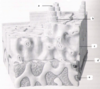
Cancellous/spongy bone tissue
- Many spaces
- Latticework of thin columns of bone (trabeculae)
- Makes up most of short, flat and irregular bones
- Epiphyses of long bones/medullary cavity of diaphyses
- Trabeculae arranged around lines of stress
- Lighter than compact bone
- Supports red bone marrow
Histology – compact bone
- Osteons (Haversian systems)
- Transverse perforating (Volkmann’s) canals
- Longitudinal central (Haversian) canals
- Concentric lamellae (calcified matrix)
- Lacunae (containing osteocytes)
- Canaliculi (osteocyte projections / communication / nutrients)

a – compact bone
b – spongey bone
c – medullary cavity
d – periosteum
e – interstitial lamellae
f – outer circumferential lamellae
g – concentric lamellae
h – blood vessels
i – lymphatic vessel
j – canaliculi
k – lacuna
l – osteocyte
m – periosteal vein
n – periosteal artery
o – outer fibrous layer of periosteum
p – inner osteogenic layer of periosteum
q – central canal
r – perforating canal
s – compact bone
t – lymphatic vessel
u – spongy bone
v – inner circumferential lamellae
w – trabeculae
x – medullary cavity
Histology – cancellous bone
- Trabeculae (no osteons or canals)
- Osteocytes in trabecular lacunae with canaliculi
- Osteocytes comparatively superficial and receive nourishment directly from blood circulating in medullary cavities

a – enlarged aspect of spongey bone trabeculae
b – details of a section of a trabecula
c – trabeculae
d – space for red bone marrow
e – lacuna
f – interstitial lamellae
g – canaliculi
h – osteoblasts aligned along trabeculae of new bone
i – osteoclast
j - osteocyte
