Blood and Cardiovascular Flashcards
What are the functions of the cardiovascular system?
Transport gases, nutrients and regulatory molecules
What are the two parts of homeostasis?
Thermoregulation and pH regulation
How does the cardiovascular system aid in protection?
Via coagulation for vascular integrity and transport of immune cells.
What are some of the characteristics of an erythrocyte (RBC)?
Transports O2
No organelles
Mainly contains hemaglobin
Spleen removes them
Last 4 months
Label the parts


Label the parts


What are the 5 types of Leukocytes?
Neutrophil: Contains specific granules
Eosinophils: Contains specific granules
Basophils: Contains specific granules
Lymphocytes: No specific granules
Monocytes: no specific granules
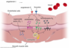
What is this and what does it do?

This is a neutrophil and it:
Is the most common leukocytes
Functions - migration to sites of inflammation, phagocytosis of bacteria, exocytosis of enzymes that attack microorganisms, results in formation of pus (dead neutrophils and cell debris)
Life span: 12-14 hours
What is this and what does it do?

This is an eosinophil and it:
Functions - migration to sites of infection and release of cytotoxins that combat parasites (protozoa, helminths), migration to sites of allergic inflammation, phagocytosis and endocytosis of antigen-antibody complexes
What is this and what does it do?

This is a basophil and like mast cells it is:
Specific granules (basophilic) – histamine (increases vascular permeability), heparin (anticoagulant), heparan sulfate, chemotactic factors for other granulocytes
Functions - similar to mast cells, upon stimulation it releases preformed and newly synthesized secretory products that initiate, maintain, and control inflammation
What is this and what does it do?

This is a monocyte and it:
Circulates 12 to 96 hours, then enters CT and differentiates
Derivatives - macrophage (Kupffer cell in liver, alveolar macrophage in lung), osteoclast in bone marrow, Langerhans cell in skin (antigen presenting dendritic cell)
Can be considered a transient cell.
What is this and what does it do?

It is a lymphocyte and there are:
B lymphocyte - humoral immunity (~20-30%)
T lymphocyte - humoral and cell-mediated immunity (~60-80%)
Natural killer (NK) cell - cell-mediated immunity (~5-10%)
Agranulocyte - lysosomal acid hydrolases
Life span: many years
What is this and what does it do?

This is a platelet and it is responsible for clotting. It has no nucleus, but has mitochondria, and vesicles with clotting factors. Binds to the injured vessel wall. Lives about 10 days.
What are the various types of Hematopoietic stem cells and what are the characteristics?
- TotipotentCapable of giving rise to all cell types (e.g., blastomere)
- Pluripotent hematopoietic stem cell (HSC)Undifferentiated cell producing blood cells of all lineages, capable of self-renewal
- Multipotent HSCUndifferentiated cell producing cells of multiple lineages, limited self-renewal (e.g., myeloid SC, lymphoid SC)
- Unipotent Committed progenitorUndifferentiated cell capable of producing cells of one lineage, colony forming units (CFUs) (e.g., erythroid CFU, granulocyte-macrophage CFU)
Label the parts


What is this? What are the characteristics?

- Cardiac myocytes are short, thick, mono- or bi-nucleated cells that often branch.
- Striated muscle cells. Sarcomere structured identical to skeletal muscle.
- Cells are attached end-to-end by Intercalated Disks. These appear as particularly dark cross-striations in cardiac muscle (arrows).
- Myocytes are not individually innervated. No somatic motor innervation.
- Gap junctions allow for functional connection between myocytes. This causes cardiac myocytes to function as a syncytium.
Where are purkinje fibers located?
In the subendocardium
Compare and contrast the TT and SR of cardiac muscle and skeletal muscle?
- T-tubules of heart are much larger in diameter than skeletal muscle.
- T-tubules do not form triads with sarcoplasmic reticulum; but instead form Diads with the more sparse cardiac sarcoplasmic reticulum.
- Cardiac T-tubules align with Z-lines, therefore number 1 per sarcomere (vs. 2 per sarcomere in skeletal muscle).
What are the parts of the arterial tunica interna?
Endothelium
Basemene Membrane
Internal elastic lamina
What are the parts of the venous tunica interna?
Endothelium
Basement Membrane
***No Elastic Lamina
What are the parts of the arterial tunica media?
Smooth muscle
External Elastic Lamina
What are the parts of the venous tunica interna?
Smooth Muscle
What are the parts of the arterial tunica adventitia?
Dense Irregular CT
Collagen Type I fibers
Vascular - only vascular layer.
Label the parts