Block 1 Flashcards

1
Q

what is the extent of the abdominal cavity?

A

Diaphragm to pelvic girdle

2
Q

Abdominal surface anatomy can be divided into 4 quadrants, what are they?

A

RUQ LUQ RLQ LLQ

3
Q

What organs sit in the RUQ of the abdomen?

A
  • colon (ascending, hepatic flexure),
  • duodenum (parts 1-3),
  • gall bladder,
  • biliary tree,
  • IVC,
  • pancreas (head + neck),
  • pylorus,
  • right kidney,
  • ureter
  • suprarenal gland
4
Q

What organs sit in the LUQ of the abdomen?

A
  • colon (descending, splenic flexure),
  • duodenum (part 4),
  • left kidney,
  • ureter
  • suprarenal gland,
  • pancreas (body + tail),
  • spleen,
  • stomach,
  • jejunum + ileum
5
Q

Which organs sit in the RLQ of the abdomen?

A
  • colon (caecum, appendix + ascending),
  • IVC,
  • right ductus deferens/ovary,
  • uterine tube,
  • ureter
  • ileum
6
Q

Which organs sit in the LLQ of the abdomen?

A
  • colon (descending + sigmoid),
  • left ductus deferens/ovary,
  • uterine tube,
  • ureter,
  • jejunum,
  • ileum
7
Q

Where is the transpyloric plane and how is it located?

A

L1

  • located halfway b/w suprasternal notch of manubrium + upper border of pubic symphysis. Passes through pylorus
8
Q

Where is the subcostal plane and how is it located?

A

L3

sits under ribs

9
Q

Where is the Supracristal plane and how is it located?

A

L4

top of iliac bone, useful landmark plane for lumbar puncture

10
Q

Where is the transtubercular plane and how is it located?

A

L5

  • corresponds to a line uniting the two tubercles of the iliac crests
11
Q

How many of each vertebrae are there?

A
  • 7 cervical vertebra
  • 12 thoracic vertebra
  • 5 lumbar vertebra
  • 5 sacral (fused) vertebra
  • 4 coccygeal (fused) vertebra
12
Q

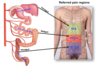
What is the point of using the regional abdomen model?

A
  • Regional models provide a standardised system for positioning/descriptions
13
Q

What are the 9 regions of the regional abdominal model?

How are the regions divided?

A
  • Split into 9 regions by mid-clavicular lines, subcostal plane (L3) + transtubercular plane (L5)
14
Q

What organs are located in the right hypochondrium?

A
  • Diaphragm
  • Costodiaphragmatic recesses
  • Liver
  • Hepatic flecture
15
Q

What organs are located in the epigastric region?

A
  • Stomach
  • Liver
  • Gallbladder
  • Transverse colon
  • Lesser sac
  • Abdominal aorta
  • Duodenum
  • Pancreas
  • Kidneys
  • Suprarenal glands
  • Origin
  • Plexus of colonic trunk
  • Superior mesenteric artery
16
Q

What organs are located in the left hypochondrium?

A
  • Diaphragm
  • Costodiaphragmatic recess
  • Stomach
  • Spleen
  • Pancreas tail
  • Splenic flecture
17
Q

What organs are located in the right flank/lumbar?

A
  • Ascending colon
  • Small Intestine
18
Q

What organs are located in the umbilical region?

A
  • Small intestine
  • Root of mesentry
  • Abdominal aorta
  • Inferior mesenteric artery origin and plexus
19
Q

What organs are located in the left flank/lumbar region?

A
  • Descending colon
  • Small intestine
20
Q

Which organs are located in the right iliac fossa?

A
  • Caecum
  • Appendix
21
Q

What organs are located in the pubic region?

A
  • Small intestine
  • sigmoid colon
  • upper rectum
  • Ovary
  • Uterine tubes
  • common iliac arteries
  • (distended bladder)
  • (Enlarged uterus)
22
Q

What organs are located in the left iliac fossa?

A

Sigmoid colon

23
Q

Which organs/pathology can refer pain to the Right hypochondrium?

A
  • Liver abscess
  • Hepatitis
  • Gallbladder/Biliary tree
  • Cholecystitis
  • Cholelithiasis
24
Q

Which organs/pathology can refer pain to the Epigastric region?

A
  • Foregut pain
  • Aortic aneurysm
  • Pancreatitis
  • Ulcer
  • Gastritis
  • Reflux
  • Myocardial Infarction
  • Pericarditis
25
Which organs/pathology can refer pain to the Left hypochondrium?
* Constipation * Splenic infarct * Diverticulitis * Pyelonephritis
26
Which organs/pathology can refer pain to the Right flank/Lumbar?
* Ascending colitis * Nephrolithiasis * Pyelonephritis
27
Which organs/pathology can refer pain to the Umbilical region?
* Midgut pain * Enteritis * Intestinal Obstruction * Mesenteric occlusion
28
Which organs/pathology can refer pain to the Left Flank/Lumbar region?
* Descending colitis * Nephrolithiasis * Pyelonephritis
29
Which organs/pathology can refer pain to the Right iliac fossa?
* Appendicitis * Gonadal pathology * Inguinal hernia
30
Which organs/pathology can refer pain to the pubic region?
* HIndgut pain * Uterine pathology * UTI/Urinary tract obstruction * Endometriosis * Pelvic inflammatory disease
31
Which organs/pathology can refer pain to the left iliac fossa?
* Diverticulitis * Gonadal pathology * Inguinal hernia * Ulcerative colitis
32
Define hernia?
protrusion of tissue/organ through a retaining tissue
33
Name some predisposing factors to abdominal wall hernias?
* FHx * loss of tissue strength + elasticity (direct inguinal hernia) * residual embryological channels (congenital hernia) * increased intra-abdominal pressure e.g. obesity, chronic constipation, chronic cough, smoking (causing chronic cough) * previous surgery
34
From superficial to deep name the layers of the abdomen
1. Skin 2. Fatty subcutaneous tissue CAMPERS FASCIA 3. Deep membraneous subcutaneous tissue SCARPA FASCIA 4. EXTERNAL OBLIQUES 5. INTERNAL OBLIQUES/RECTUS ABDOMINIS 6. TRANSVERSUS ABDOMINIS 7. TRANSVERSALIS FASCIA 8. Extraperitoneal fat 9. PARIETAL PERITONEUM
35
What are the muscles of the abdomen? What direction are the muscle fibres?
* External Oblique * Internal Oblique * Rectus Abdominis * Transversus Abdominis
36
What are the functions of rectus abdominis? What happens to rectus abdominis during pregnancy?
1. Flexing the torso + vertebral column in the abdominal region 2. Compression of the abdomen 3. Stabilisation of the torso **Divarication of rectus abdominis muscles (DRAM)**/diastasis recti --\> right + left sides of the rectus abdominis spread apart at the stomach midline (linea alba) --\> can lead to hernia formation around the umbilicus
37
What is the linea alba?
White line --\> midline fibrous aponeurosis Attachments of linea alba: * xiphoid process * pubic symphysis * aponeuroses of abdominal muscles Linea alba is relatively **avascular** + **aneural** therefore it is a good access point for abdominal surgery --\> blood loss is minimal + major nerves are avoided _Can insert needle/cannula safely through linea alba_
38
Describe the location of the external and internal oblique muscles
* **External oblique** --\> superficial to internal oblique, passes inferomedially from T5-12 to linea alba, iliac crest, anterior superior iliac spine (ASIS), pubic tubercle * **Internal oblique** --\> deep to external oblique, lower fibres pass medially + upper fibres pass superomedially from the throracolumbar fascia, iliac crest + lateral inguinal ligament to the linea alba, lower ribs, upper pubis + conjoint tendon
39
Where is the Inguinal ligament?
* the free lower border of external oblique passing from anterior superior iliac spine (ASIS) to pubic tubercle
40
What lies between **transversus abdominis** and **internal oblique** muscles?
A neurovascular plane
41
What is the rectus sheath formed of? what does it enclose? What is the significance of the arcuate line?
* The **rectus abdominis** muscle is contained within a fibrous sheath formed by the aponeurosis of **transversus abdominis**, **external oblique** + **internal oblique** muscles + transversalis fascia Rectus sheath encloses rectus abdominis + epigastric vessels --\> _inferior epigastric artery + inferior epigastric vein_ (blunt-force trauma can cause bleed + increase intra-abdominal pressure resulting in pain --\> **rectus sheath haematoma**
42
Name the collective functions of the muscles of the abdominal wall
* Support * Protection * Posture maintenance * Intra-abdominal pressure to support defecation, micturition + parturition (also vomiting + coughing) * (_Weakness of part of the abdominal wall musculature can result in **hernia**_)
43
44
What are the Attachements, Innervation and Function of the Internal Obliques
Attachments: Originates from ribs 5-12, and inserts into the iliac crest and pubic tubercle. Functions: Contralateral rotation of the torso. Innervation: Thoracoabdominal nerves (T7-T11) and subcostal nerve (T12).
45
What are the attachments, function and innervation of the Internal Obliques?
Attachments: Originates from the inguinal ligament, iliac crest and lumbodorsal fascia, and inserts into ribs 10-12. Functions: Bilateral contraction compresses the abdomen, while unilateral contraction ipsilaterally rotates the torso. Innervation: Thoracoabdominal nerves (T7-T11), subcostal nerve (T12) and branches of the lumbar plexus.
46
Describe the course of the superior and inferior epigastric arteries
* ![]()**Superior + inferior epigastric arteries** travel + meet in the rectus sheath --\> unite the subclavian artery with the external iliac artery --\> forms an arterial shunt if the aorta is narrowed
47
Explain the motor nerve supply of the abdominal muscles and how would a patient present with damage?
* carries nerve impulse from CNS which trigger muscles to contract * _damage could cause weakness, twitching + paralysis of abdominal muscles in this region (could result in hernia)_
48
Explain the sensory nerve supply of the abdominal muscles and how would a patient present with damage?
* carries sensory information towards the CNS * _damage could cause pain, numbness, tingling, increased sensitivity, burning + problems_
49
Why are certain surgical incisions used?
* Surgical incisions/endoscopy ports take into account the position + course of arteries + nerves in order to minimise iatrogenic damage * Incisions are based upon the organ/region to be accessed + route of minimal damage * Increasing use of endoscopic access has led to these approaches being used less frequently
50
What are the names of these surgical incisions? What is the relevance of them?
MEDIAN/MIDLINE: linea alba incised. Tissue is relatively avascular - slow healing PARAMEDIAN: Recust sheath incised, rectus muscle displaced laterally to avoid nerve damage GRIDIRION (muscle splitting) at MCBURNEY'S POINT: What nerve is at risk? potential consequence PFANNENSTIAL (SUPRAPUBIC): what nerves are at risk? SUBCOSTAL (KOCHER): Inferior to costal (thoracic) margin - what are at high risk of damage?
51
Outine the lymphatic drainage of the abdominal wall
Abdominal wall lymphatic drainage passes to axillary and inguinal nodes. Lymph drainage follows quadrants: * upper quadrants drain to axillary nodes * lower quadrants drain to superficial inguinal nodes The lymphatic drainage of more superficial tissues + skin is regional
52
What are lymph nodes?
Lymph nodes : small swellings of lymphoid tissue in the lymphatic system where lymph is filtered + lymphocytes are formed. Part of the immune system
53
What is lymphatic fluid?
Lymphatic fluid: clear/white fluid that is collected, filtered + transported by the lymphatic systems from around the tissues to the blood circulatory system. Contains lymphocytes
54
Where is the gut tube located during development?
* The gut tube is located within the peritoneal cavity + is surrounded by a layer of tissue called peritoneum (serous membrane)
55
Explain what is meant by : intraperitoneal Retro-peritoneal Secondary retro-peritoneal Mesentry
**Intra-peritoneal** --\> structure covered in peritoneum **Retro-peritoneal** --\> structure behind (outside) of the peritoneum **Mesentery** --\> double-layered fold of peritoneum suspending an organ from the abdominal wall **Secondary retro-peritoneal** --\> intra-peritoneal structure that later becomes retroperitoneal
56
The gut tube blood supply arises from 3 main arteries which branch from the abdominal aorta, what are ther?
**Coeliac trunk** (T12) supplies **foregut** **Superior mesenteric artery** (L1) supplies **midgut** **Inferior mesenteric artery** (L3) supplies **hindgut**
57
What does the foregut consist of?
**(lower oesophagus --\> major duodenal papilla)** lower oesophagus, stomach, spleen, liver, gall bladder, pancreas, 1st + 2nd part of duodenum
58
What does the midgut consist of?
**major duodenal papilla --\> last third of transverse colon** 3rd + 4th part of the duodenum, jejunum, ileum, caecum, ascending colon, transverse colon (2/3)
59
What does the hindgut consist of?
**last third of transverse colon --\> upper anal canal** transverse colon (1/3), descending colon, sigmoid colon, rectum, upper anal canal
60
How is visceral pain felt?
vague, diffuse + poorly defined/located sensation
61
What are viscera sensitive to? What are viscera not sensitive to?
Sensitive to stretch, hypoxia, chemicals & environmental changes Not sensitive to cutting or thermal stimuli
62
What is a dermatome?
an area of skin innervated by a single spinal nerve (this is the evidence-based dermatome to which we standardise
63
Describe why visceral pain is vague and poorly defined
Visceral (organ) + somatic sensory (afferent) nerves enter the spinal cord together + travel in the same spinal tracts 1. Brain confuses location/origin of signal 2. Perceives that pain is of dermatomal (skin) origin 3. Sensory nerves from organs share pathways/routs of travel with other sensory nerves
64
Outline regions of referred paim
65
What are visceral nerves?
* autonomic division of NS, conduct impulses to + from internal organs, glands + blood vessels
66
What are somatic nerves?
part of peripheral NS associated with the voluntary control of body movements via skeletal muscles
67
What are sympathetic nerves?
part of autonomic NS that prepares the body to react to stresses such as threat or injury (e.g. fight or flight). Short neurones, quick response
68
What are parasympathetic nerves?
part of autonomic NS that controls functions of the body at rest + helps maintain homeostasis in the body
69
What are afferent nerves?
* nerve that carries sensory nerve impulses from the periphery towards the CNS
70
What are efferent nerves?
* nerve that carries impulses from the CNS toward the periphery e.g. motor nerve
71