Bioenergetics Flashcards
Reference reading for bioenergetics
REFERENCE READING: Medical Biochemistry. An Illustrated Review. Panini, SR. Thieme, c2013, Pages 127 -148.
- Disorders of the Krebs cycle – see supplied PDF (reading material-highlighted sections)
- Genetic alterations in Krebs cycle and its impact on cancer pathogenesis – see supplied PDF (reading material-highlighted sections)
Explain the importance and regulate of pyruvate dehydrogenase complex (PDC)
Phosphorylated form of PDC is inactive • Phosphorylation occurs in coenzyme Thiamine pyrophosphate (TPP) of E1 complex • Dephosphorylated version of PDC is active
In a phosphatase deficiency PDC is always in phosphorylated form (inactive) Glucose Lactate rather than Acetyl CoA Results in constant lactic acidosis (i.e., high blood levels of lactic acid) Central nervous system is effected the most Alanine intake should be restricted
(p. 128‑129; Table 10.1, Fig. 10.2)
Explain PDC in tissues
Correlation box
Explain Pyruvate dehydrogenase deficiency (neonatal lactic acidosis)
correlation box (p. 129)
What is the significance of Arsenite and lipoic acid?
correlation box (p. 129)
Describe Beriberi and Wernicke-Korsakoff Syndrome [Students should comprehend the importance of thiamine in the function of PDC]
correlation box (p. 129)
Compare and contrast the TCA cycle reaction steps and identify where high-energy equivalent substances are formed
Steps 1 and 2 : Condensation and isomerization to generate isocitrate Step 3 and 4 : Irreversible oxidation and decarboxylation NADH, CO2 , and Succinyl CoA Step -5: Formation of Succinate and GTP Step 6 -8: Produce FADH 2 , NADH, and regenerate Oxaloacetate Steps 1,3, and 4 : Regulated steps
When cellular ATP levels are low, the activity of TCA cycle is increased High levels of cellular ATP triggers the inhibition mechanism of TCA cycle mitochondrial ETC inhibition
(Fig. 10.3, p. 130-131)
Describe Differential effects of citrate on rate-limiting enzyme
correlation box (p. 130)
What are the ATP:ADP and NADH/NAD+ ratios?
correlation box (p. 132)
How does rat poison inhibit the TCA cycle?
correlation box (p. 133)
Describe succinyl CoA in heme synthesis
(p. 133)
Compare and contrast the anaplerotic reactions and anabolic functions of TCA cycle. Describe how the TCA cycle switches to make intermediates in fasting and fed state.
• Anaplerotic “fill up” reactions provide intermediates for replenish TCA Cycle • Two major anaplerotic reactions 1. Degradation of amino acids 1. Carboxylation of Pyruvate
(p. 133‑135; Fig. 10.4; Fig. 10.5 A-B)
Describe the effects of Pyruvate carboxylase deficiency
(p. 134)
Discuss disorders of TCA cycle
Mitochondrial depletion syndrome associated with Profound hypotonia Progressive dystonia Muscular atrophy Severe sensory neural hearing impairment
(see reading reference)
Describe Oxoglutaric acid aciduria
2-Oxoglutaric aciduria (a-ketoglutaric acid) A rare disorder with global developmental delay and severe neurological problems in infants Metabolic acidosis Severe microcephaly Mental retardation
reading reference
What happens in Fumarase deficiency?
Characterized by severe neurological impairment. Fatal outcome within the first 2 yrs. of life Encephalomyopathy Dystonia Increased urinary excretion of fumarate, succinate, aketoglutarate, and citrate Autosomal recessive disorder Mutation in fumarase gene contains Q319E
reference reading
Succinyl CoA synthetase deficiency
Succinyl-CoA synthetase (SCS) deficiency Associated with mutations two out of three subunits making up the enzyme, SUCLA2 and SUCLG1 These genes encode the β-subunit of the ADP-forming SCS and α-subunit of SCS Oncometabolites of TCA cycle are Citrate and 2-hydroxyl glutarate
reference reading
What are the Oncometabolites of the TCA cycle?
Oncometabolites are small biomolecules (or enantiomers) of normal metabolism. Excessive accumulation of them causes metabolic dysregulation [91]. Consequently, cells with high oncometabolite concentrations undergo progression to cancer [91]. Oncometabolites of the TCA cycle are accumulated due to mutations of genes involved in metabolism.
The oncometabolites can be divided in two categories - (I) native oncometabolites that could be accumulated due to mutations (loss of function) of genes encoding the enzymes, and (ii) promiscuous oncometabolites that could be accumulated due to gain-of-function mutations in metabolic enzymes.
reference reading
Compare and contrast free radicals as oxidizing agents, signaling molecules, and cellular response to invading microorganisms
(Fig. 12.18, p. 194)
Define the respiratory chain and discuss the mechanisms of OxPhos and Components of respiratory chain
A successful OxPhos must accomplish the following key goals : 1. To transfer electrons from NADH and FADH2 O2
- To establish a proton gradient across the inner mitochondrial membrane and in intermembrane space
- To synthesize ATP
(p. 138; Fig. 11.2; p. 140-143) AND (Table 11.1)
Cytochrome-c and apoptosis
correlation box (p. 137)
What are the Respiratory chain components encoded by mtDNA?
correlation box (p. 144)
What is Rotenone?
correlation box (p. 144)
Cyanide poisoning
correlation box (p. 144)
Describe Cyanide versus carbon monoxide action
correlation box (p. 144)
Describe Aspirin overdose
correlation box (p. 145)
What is Electron transfer?
1)-Transfer of Electrons The electron flows from the molecules with lower E0 ’ to that with highest E0 ’ The difference in Δ E0 ’ is associated with ΔG0’ Δ E0 ’ and ΔG0’ are inversely related ΔG0’ = -nƑ Δ E0 ’
Inhibition of electron transport • When transfer of electrons inhibited A decrease in the pumping protons A decrease in the proton (H+) gradient Inhibition of ATP synthesis
reference reading
Proton gradient and proton motive force (Δ pmf)
Electron transfer through the respiratory chain lead to the pumping of H+ from matrix to the innermitochondrial space Two factors constitutes a proton-motive force (pmf) to drive ATP synthesis by Complex V 1) pH gradient (ΔpH) 2) Membrane potential (ΔΨ)
reference reading
Chemiosmosis theory- Compare and contrast coupling and uncoupling of proton gradient and is consequences.
Uncoupling [disruption of protein gradient] In OxPhos Electron transfer couples to proton gradient P ~ADP ATP
If the proton gradient is disrupted P ~ ADP uncouples from the electron transfer Protons (H+) reenter the mitochondrial matrix from the intermembrane space TCA cycle and electron transfer to O2 are accelerated ATP synthase is inhibited (No ATP Synthesis) Heat generation
(p. 142; Fig. 11.6).
Compare and contrast the mitochondrial electron transport chain (ETC) toxins
(p. 142; Fig. 11.6)
Synthesis of ATP
3)- Synthesis of ATP • Catalyzed by a large membrane-bound protein • ATP synthase (Complex V) • Harnesses the energy contained in pmf, obtains necessary power (7.3 kcal/mol) to form ADP + Pi ATP Inhibitors: Oligomycin disrupts proton transport through the channel
reference reading
Regulation of OxPhos
Generates most of the ATP It is sensitive to O2 [ATP/ADP] ratio
(p. 143; Fig. 11.7)
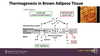
Thermogenesis in brown adipose tissue
(p. 144)
Other electron transport systems
correlation box (p. 145)
Hypoxia and ATP preservation
correlation box (p. 145)
Nucleotide analogue treatment and fatigue
correlation box (p. 145)
Antioxidants and removal of free radicals
Antioxidants • Superoxide dismutase (SOD) O-. 2 + 2H+ O2 + H2O2 • Catalase 2H2O2 O2 +2H2O • Glutathione peroxidase • Vit-E • Vit-C
Superoxide dismutase 1. Cu/Zn SOD (SOD1) cytosolic 2. Mn/Zn SOD (SOD2) mitochondrial
reference reading
Free radicals and antioxidants
Correlation box (p. 10)
Ubiquinone radical
Correlation box (p. 145)
Explain the mitochondrial transport system. Compare and contrast tissue specific shuttle systems
(p. 146-148).
Malate-Aspartate shuttle system
Malate-aspartate shuttle Operates in the heart, liver, and kidneys Generates NADH in mito-matrix NADH enters to ETC at Complex-I
(p. 147; Fig. 11.8 A)
Glycerophosphate shuttle system
Glycerophosphate-shuttle Operates in skeletal muscle and brain Generates FADH2 in the inner mitomembrane FADH2 joins to ETC at CoQ
(p. 147; Fig. 11.8 B)
Antiporters for phosphate / OH¯ and phosphate / malate exchange
(p. 148; Fig. 11.9 A, B)
Antiporter for ADP / ATP exchange
(p. 148; Fig. 11.9 C)
Antiporter for pyruvate / OH¯
(p. 148; Fig. 11.9 D)
Sources of NADH and FADH2
Correlation Box (p.145)
Other electron transport systems
Correlation Box (p. 145)
Discuss mitochondrial medicine: An overview
reference reading
First mitochondrial disorder; Luft’s
disease reference reading
Causes of mitochondrial disease
• Primary cause : – Defect in nuclear DNA (nDNA) encoding the mitochondrial proteins – Defect in mitochondrial DNA (mDNA) • Secondary causes: – Ischemia – Reperfusion – Cardiovascular diseases – Renal failure – Drugs and aging – Alcohol – Smoking. – Others
reference reading
Defect in nuclear DNA (nDNA)
reference reading
Defect in mitochondrial DNA (mDNA)
reference reading
Manifestations of mitochondrial disease
Manifestations of mitochondrial diseases
- Clinical features – Nervous system: seizures,ataxia,dementia, deafness, blindness – Eyes : ptosis, external ophtalmolplegia,retinis pigmentosa with visual loss – Skeletal muscle : Muscle weakness, fatigue, myopathy, exercise intolerance, loss of coordination and balance – Heart: cardiomyopathy – Others: gastrointestinal, liver failure, kidneys, pancreatic disease, diabetes, etc.
- Metabolic Features – Low energy production – Increased free radical production – Lactic acidosis
reference reading
TCA cycle and cancer
Studies demonstrated that abnormalities of the TCA cycle are associated with different types of cancers [25e27] (Fig. 1). For example, it was noted that phosphoenolpyruvate carboxykinase, a regulator of TCA cycle flux, plays a crucial role in promoting cancer cell growth and proliferation, particularly in colorectal cancer cells [27]. Phosphoenolpyruvate carboxykinase increases glucose and glutamine uptake in cancer cells and favours anabolic metabolism [27]. This metabolic shift is important for highly proliferating cells, like cancer cells, which continuously require a supply of precursors for the synthesis of lipids, proteins and nucleic acids [28]. Several studies also demonstrated that dysfunction of mitochondrial metabolic pathways such as the TCA cycle and oxidative phosphorylation are associated with the conversion of non-neoplastic mammary epithelial cells to non-metastatic breast carcinoma and then to the metastatic cancer cells [29e31].
Describe the role of excess Citrate in cancer.
Excess citrate reduces the activity of the mitochondrial isoform of pyruvate dehydrogenase and results in a shift of the cell’s metabolism towards glycolysis [94]. Increased accumulation of pyruvate leads cells to convert pyruvate to lactate and regenerate NAD þ which is the key factor for glycolysis [94] (Fig. 1). Thus citrate accumulation further favours the non-oxidative breakdown of glucose in the cells and promotes cancer growth.
Increased accumulation of citrate activates the enzyme acetyl CoA carboxylase (ACC), which in turn increases the production of acetyl CoA and malonyl CoA [97,98]. This increased acetyl CoA and malonyl CoA is then directed towards increased lipid/sterol synthesis [97,98]. The transformed lipid metabolism observed in cancer cell shifts the metabolic fate of lipid synthesis of the cell membrane and changes the redox potential of cells. These alterations promote cellular processes like cell growth, proliferation, cell survival signalling and differentiation which in turn contributed in the tumorigenesis [99].
Describe the role of 2-Hydroxy glutarate (2HG) in cancer
. 2-Hydroxy glutarate (2HG) Mutations of IDH1 and IDH2 (cytosolic and mitochondrial isoforms of IDH) lead to the accumulation of 2HG and acts as a pathogenic metabolite (oncometabolite) in many cancers [42,52,106e110]. A study also reported that this mutation was involved in the conversion of alpha-ketuglutarates to R (-)-2-hydroxyglutarates (2HGs) [41]. Excessive accumulation of these 2HGs acts as an oncometabolite and led to the malignant progression of gliomas
