BIoChem Mini 3 Flashcards
Palmitic Acid (16C)
- Primary fatty acid
- Modification of this molecule produces other types of fatty acids FAs.
- The body first makes this fatty acid and then modifies it into other FAs
- Can only add double bonds up to C9 in mammalians.
Pancreatic Pseudocysts
marker is very high level of alpha-amylase (in the thousands) -noted by fluid filled cysts on the pancreas contaianing amylase, lipase and zymogen, etc.

Statin Drugs (What, How?)
- -Obtained from fungal source -inhibits HMG-COA-reductase and endogenous cholesterol biosynthesis ( lower total Chol and LDLs)
- -Lowers intracellular cholesterol concentration -increases LDL biosynthesis and promotes LDL clearance
* lowest total cholesterol levels but long-term usage have side effects such as muscle pain and cramps due to deficiencies of isopreniods
Pyruvate Carboxylase Deficiency Disease
- N.Amer Type: enzyme produced but nonfunctional; severe mental/DD
- UK & France: nonsense mutation in gene. NO Enzyme at all. Death in infants (3-months)
- Benign: quantity lacking in enzyme. Metabolic acidosis
Alpha-1 Antitrypsin/Alpha-1 Antiprotease
- Made in Liver
- Acute Phase Reactant
- Inhibits Trypsin & Elastase (prevent break down of connective tissues by elastase)
- multiple gene variant that can lead to liver & Lung diseases.
- Z-Allele ( gene variant) can lead to neonatal hepatitis due to accumulation in the hepatocyte ER.
Creatine Kinase (CK)
Creatine Kinase helps to phosphorylate creatine and store it in muscles. NORMALLY ALMOST ABSENT IN BLOOD PLASMA
- Expressed in muscle cells
- CK consist of two protein subunits; combine to form 3 isoenzymes. Can be separated by Electrophoresis.
- M (for muscle)
- B (for brain)
- BB (CK1): expressed primarily in brain; elevated plasma levels this marker for brain damage
- MB (CK2): expressing a cardiac muscles; elevator plasma levels market for cardiac muscle damage
- MM (CK3): found in skeletal and muscles. elevator plasma levels marker for muscular dystrophy

Gamma Glutamyl Transaminase (GGT)
Diagnostic marker for liver, kidneys, pancreas, and prostate cells
- Levels are increased in Males
- Increased GGT levels and blood due to alcohol and drug effects on liver.
- Elevated GGT and AST levels are markers for alcoholic liver cirrhosis
(AST/ALT) Ration>1
Wilson’s Disease
Low cerulupllasmin in the blood and abnormal levels of
Autorecessive disorder associated with copper metabolism due to defective ATP7B.
- Absoption of copper in intestine is okay but once it is n the liver cell; binding of the copper doesn’t happen.
- Toxic buildup of copper, leaks into blood.
- Abnormal deposit of copper in the organs (brain, Liver, eyes, lungs)
- show neuro, mental, and rickett like symptom, kidney disorders, cardiac, and eye issues (kayser-feleischer rings).
Ceruluplasmin (Alpha-2 Globlin)
Liver Glycoprotien: Copper containing; helps to t/p copper in blood and distribute. Liver helps to maintain copper balance/regulation by hepatocytes. (ATP7B attaches).
- Absopted in intestine
- Func: Cytochrome C, need to produce ATP.
- Binds to 6 cupper atoms
- Helps to oxidize iron and t/p.
- (10% of copper is t/p by albumin)readily aval
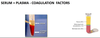
Serum & Plasma
( what are they and where are they made)
Contains both simple and conjugated proteins (glycoprotiens and lipoproteins)
- Synthesized in the liver.
- Serum is yellow are is the top part left after the blood clots in the test tube.

Aspartate Transaminase (AST)
Diagnostic marker for Liver.
- Present in cytoplasm and mitochondria of hepatocytes. Also present in muscles and cardiac muscles.
- transfers AA-ketoacid
–(AST/ALT) Ratio slightly
–
Hemoglobinuria
Hemoglobinuria: Hb appears in urine
Can lead to Kidney damage
Antitrypsin Deficiency
- Can lead to Emphysema due to neutrophil elastase remain active in the lungs mediating lung damage.
- Smoking can lead to emphysema b/c H202 oxidizing methionine in antitrypsin turning it into methionine sulfoxide.
Menkes Syndrome
Genetic disorder (X-linked)
Defective ATP7A protein that affects dietary copper form intestine enterocytes via blood.
- Copper builds up in the intestine & kidney.
- Copper deficiency in brain and other organs
- Structure of bone, skin (scaly), hairy (kinky) and blood vessels affected.
- Affects nervous tissue function.
family HX, hair, lack of copper, sex- Vignette keys.
Monoclonal Gammopathy
Abnormal Amount of one type of Ab in the blood
2Types:
- Multiple Myeloma
- Waldenstrom’s Macroglobulinemia
Biotin Carrier Protein (function?)
Attaches carbonyl group to biotin
(ALP) Alkaline Phosphotase
Marker for Liver and Bones Diseases Marker for
- Dephosphorylates proteins, nucleic acids etc. -Levels higher in children, 3rd trimester, and elderly.
- found in biliary ducts and osteoblasts
Liver
Cholestasis (bile duct obstruction, gallbladder tumors etc)
Bone
Osteomalacia/Rickets: Elevated ALP &BAP
Heptogloblin
(alpha-globlin)
Carries Hemogloblin dimer release in blood to liver/macrophages for degradation.
- Glycoprotein sysnthesized in the liver
- Globlin & iron of Hb are recycled; heme is metabolized into bilirubin & excreted.
- Free hemogloblin (hemogloblemia) is usually due to infection or hemolysis.
Plasma Protein Electrophoresis
Separate proteins in mixture based on size and charge density.
Mobility of proteins are based on charge and size
- Smaller fragments move towards bottom
- Negative moves towards Anode (Top)
- Positive moves towards cathode (Bottom)
Albumin: Major component Forms Largest Peak close to anode (top)
Gamma Globulin: Closest to the (-) electrode/cathode (bottom)

Albumin
EDEMA Develops when albumin conc in plasma/serum drops below 2g/dL
- Major Plasma Protein content
- Single peptide chain
- Best Buffer in the Blood
- Helps with reabsorption at the tissue/cap bed level
- lightest and more negative
Relatioship between edema and colloid pressure. proteins in veins attract H20 and electrolyes which pulls fluids back into the cappillaries at the venous end.
What is the rate limiting step in FA biosynthesis?
(Rate limiting step) Acetyl CoA carboxylation via biotin to become Malonyl CoA
- Allosteric Regulation
- Citrate is a Positive Effector
- Palmitoyl CoA/Palmitic Acid are Negative Effectors

_Transferrin (TF)
(beta-globlin)_
Glycoprotien from liver
- Negative Acute Phase Protein
- Fe (Iron) metabolism/ Tranfers Iron from liver/gut to bone marrow (Rate 2Fe+3 per TF molecule)
**High in Fe deficienciency anemia
Normal in other typeps of anemia

Non-enzymatic pathway autooxidation
- Catalyzes by free radical mediation
- called lipid peroxidation
- produces hydroperoxides
- membrane cholesterol/LDL subset the boat to nonenzymatic oxidation
- so you would damage and chain reactions and also lead to atherosclerosis
Globulins
- Synthesizes alpha and beta in Liver
- Gamma Globlins ( antibodies( systhesized in the reticulo-endothelial cells ( plasma, b-Cells)
Alpha-1-antitrypsin
Troponin
Marker for MI
- Not an enzyme
- Troponin: C, I, T ( calcium binding protiens) present in the cytosol of the myoctye
- Not present in blood under normal circumstances
- Troponin I&T can present in cardiomyocytes; very important marker for MI.
Release in blood within 2-4 hrs
Peaks between 12-24 hrs
Presists in blood for up to 7-days
HIGH TROPONIN LEVEL ARE EVIDENT IN MI, PE, MYOCARDITIS.

Hypoalbuminemia
Occurs due to:
Decreased albumin synthesis
Increased catabolism of albumin
Can occur due to liver issues such as cirrhosis, nephrotic sysndrome, burns , etc.
Hypoalbuminemia leads to edema
Hemopexin
(beta-globlin)
Does same thing as heptogloblin; except it binds Heme not Hb.
- synthesized in the liver.
- Binds free Heme from intravascular hemolysis and take it to the liver to be converted to bilirubin.
- Low levels of hemoplexin during severe hemolytic anemia.
Lactate Dehydrogenase (LDH)
Found in lots of places. Converts pyruvate to lactate when in hyoxic conditions
- High Conc in cardiac and skeletal muscles
- 4 polypetide chains that give rise to 5 isoenzymes
- 2 types: H, M types
WHEN YOU SE ELEVATED LDH THAT MEANS CELL DAMAGE HAS HAPPENED.
When you see evelated LDH that means that you must order more specific markers to see what is going on.

Pancreatic Amylase & Lipase
- *Marker of Acute Pancreatitis (levels elevated 5-10 times)**
- elevated plasma levels & low urine levels of alpha-amylase suggests severe glomerular issues.
Exocrine: 3 important enzyme:
Typsin ( protease), Pancreatic alpha-amylase ( breaks down simple carbs), Lipases ( helps break down comp fats into glycerol). ( THESE 3 DIGEST PROTEINS, LIPIDS AND CARDS IN DUODENUM & JEJUNUM)
All 3 produced in inactive form until typsinogen is activated in intestines; Typsin becomes activated and then it activates Amylase and Lipase.
* if typsin is activated in pancreas it will tell auto-digest pancreatic acinar cells cause inflammation known as Pancreatitis.
Hyperproteinemia
Relative or Absolute increase in total protein conc in plasma/serum
- Relative: [Protein] results from change in [water}.
- Absolute: Occurs due to increased protein synthesis.
May occur due to increased protein loss, blood loss, low protein intake, bad burns, edema(relative).
Acute Phase Reactants
Serum Proteins that respond ( elevate/decline) to acute inflammatory conditions.
Cytochrome P450 Enzymes
- Involved in enzymatic pathway of cholesterol oxidation which is production of bile.
- Catalyzes Enzymatic pathway -
- Leads to synthesis of bile -occurs in hepatocytes
Fatty acid synthase (FAS) complex
- Catalyze the synthesis of FA Single multifocal polypeptide chain with 7 active sites
- Contains 2 carrier proteins (shuttlers)
- Acyl Carrier protein ( ACP)
- beta-ketoacyl synthase (K-SH)
Paget Disease
Metabolic bone disease due to excessive rate of bone remodeling.
- Diagnostic markers are bone alkaline phosphatase (BAP) conc elevated
Duchene Muscular Dystrophy
Affects kids, can be inability to stand with muscle weakness, in extremities.
MM (CK3): found in skeletal and muscles. elevator plasma levels marker for muscular dystrophy
MM will be greatly elevated.
Acid Phosphatase
Diagnostic marker for Prostate Cancer
- Widely avaliable in bones, liver, spleen, kidney, RBCs
- Higher levels in Males due to Prostate.
SIGNIFICANT ELEVATED: MARKER FOR METASTATIC PROSTATE CANCER
MODERATE ELEVATION: Bone disease, lysosomal disease, Sickle cell, etc.
Multiple Myeloma
Abnormal proliferation of single clone* of *plasma cells. Prevalent neoplastic disease.
- Excessive amounts/production of immunogloblins molecules.
- Hematological cancer
- IgG myelona: 50%
- IgA myeloma 25%
- Monoclonal production of light chains: 20%
- Affects Afr Amers more in US
- Survivial is 3-10 years afer Dx
Common syptoms: “Bone pain”, infections, hypercalemia, renal issues, fractures, neuropathies.
- Lytic Bone Disease
- Hematological issues: leukopenia, anemia, thrombocytopenia
- Kidney: stones and high blood Ca level =kidney dysfuction
Dx Features:
- Abnormal increase in serum protein
- Plasma serum electrophoresis ( abnormal M-Spike of M-Protien)

2 pathways for oxidation of cholesterol
- enzymatic pathway catalyzed by cytochrome P450 enzymes
- non-enzymatic pathway (auto oxidation)
ELISA (Enzyme Linked Immunosorbent Assay)
3 Ways
-Direct: Label Primary Antibody only; no secondary
-Indirect: Label Secondary, which is attachedto unlabeled Primary
-Sandwiched: same as indirect but thereis an unlabeled captured antibody.

Role of CK-2 ( MB) in MI
CK-2 level very high in response to cardiomyopathy
Take 3-6 hrs for CK-2 to be in blood
Peaks in 12-24 hours
Returns to pre-MI level within 12-24 hours
Total CK &CK-2 important for Dx of MI
Hypergammaglobulinemia
(gammopathy)
- High amt of gamma globlins. High amount of immunogloblins.
- Abnormal proliferation of plasma cells
- Monoclonal gammopathy
Types of Alpha-2 Globlins
- Heptogloblin
- Ceruloplasmin
Alanine Transaminase (ALT)
Diagnostic Marker for Liver
- Present only hepatocytes
- Available in high concentrations then AST.
(AST/ALT) Ratio slightly
(AST/ALT) Ratio
(AST/ALT)Ration>1 ( alcohol induced liver issues)
Waldenstrom’s Macroglobulinemia
Rare monogammopathy with High Levels Of IgM (pentamers) in blood.
- Clonal disease of B Lymphocyte
- Called indolent Lymphoma b/c it spreads slowl
- Common S&S: different from multiple myelnoma. Nose&gum bleeding***, ***Numbness/tingling*** in ***extemities in cold that return to normal in warm. blurry vision/HA
- Clinical Features:*
- *Hyperviscosity:**Excess IgM increases serum viscosity =circulatory impairment.
- *Anemia: Excessive** B-Cells in bone marrow hinder RBC production.
- *Cryoglobulinemia:** Single/mixed (IgM/IgG/IgA); undergoes REV precipitation at low temps. causes hyperviscosity of plasma, cap spasms, pain, cyanosis, tingling of fingers in cold weather.
(lytic bone& kidney dysfunc are not common in these Pts.)
DX: CBC, Serum Eletrophoresis ( for M-Spikes), Cryocrit ( looking for plasma cryogloblins), bone marrow biophy (abnormal # of malignant lyphocytes in bone marrow)

Primary Hemostasis
Vasoconstriction and formation of platelet plug

Secondary hemostasis
activation of coagulation cascade; stabilization of plug ( formation of fibrin network.

Tertiery hemostasis
Dissolution of the fibrin meshwork
- Clot Retraction: retraction of the edge of the wound; started by contractile proteins in platelets
- Fibrinolysis: plasmin mediated breakdown of clot.
- *tPA:** Dissolves clots used in tx strokes
- *Streptokinase:** fibrinolytic Rx used to dissolve clots by activating plasminogen into plasmin.
**Urokinase & tPA play a big role in this process.
vWF Complex
Factor VIII + VWF COMBINED
Prevents Factor VIII from binding.
Release of Factor VIII initiated by Thrombin
Factor VIII then used in Intrinsic Pathway.
Thrombocytopenia
Low platelet count (<40,000/microL) may affect platelet plug formation
Von Willebrand disease (vWD)
Clinically heterogenous hereditary hemorrhagic disease classified into the following three types:
vWDI: Symptoms. Common. Caused by a quantitative deficiency of the vWF (20-50%)
vWDII: Structural abnormalities of vWD protein ; vWF & Factor VIII binding is slowed down.
vWDIII: Rare vWF-mutation disease. TOTAL ABSENCE of vWF protien. Pts will blood badly/hemmorrhagic.
Extrinisic Pathway
Activation of multiple coagulation factors (serine protease enzymes)
starts generation of active factor Xa (central element of both cascades)
Factors: 3, 7, 10
Xa=converts Prothrombin to Thrombin which converts fibrinogen into fibrin.

Intrinsic Pathway
Amplifies generation of Factor Xa.
- Activated by contact ( implants shoudnt be charged it will cause clots)
- Activates Xa, makes large amts.
- 1 molecule of Xa can make 1000 of thrombin.
Factors: 5, 8, 9, 10, 11, 12
Xa=converts Prothrombin to Thrombin which converts fibrinogen into fibrin.

Common Steps in Extrinsic

X—> Xa

Tenase Complex
(Factors 9a & 8a)
Intrinsic Pathway
Factos IXa, VIIIa bound to platelet membrane phospholipds (CA2+ dependent).
Factor X binds to tenase complex and activates Xa
Tenase along with prothrombin complex requires Ca+
Hemophilia
X-linked recessive disorder; occurs in males; females are commly asymptomatic genotyphic carriers.
Cause: deficiency of specific coag factors
- *2 Types:**
- Hemophilia A:** Deficiency of *Factor VIII
- Hemophilia B:** Deficiency of *Factor IX (christmas disease)
Combination of A&B leads to death in newborns.
Symptoms: Abnormal bleeding. moderate= response to trauma/surgery; severe: spontaneous.
Protein C (PC)
Thrombosis regulator.
Trypsin like serine protease; major regulator of coagulation; made by liver
Protien S (PS)
Thrombosis Regulator
Cofactor of activated Protien C (PC)
Thrombomodulin
Thrombin bounds to endothelial thrombomodulin and activates PC to PCa
- aPC & PS inactivate Va, VIIIa
- Blocks formation of prothrombinase complexe
- Blocks activation of factor X
Heparan Sulphate
(short term anticoag)
Activator of Antithrombin III; potentiates the activity of antithrombin III
Mimics antithrombin III slows down the coag cascade/ short term anticoagulate.
Antithrombin III (Serpin)
Regulator of coagulation
Neutralizes thrombin; stops activitites of Factor IXa, Xa, XIa, XIIa, plasma and kallikren.
Vitamin K
Cofactor of the liver enzyme gamm glutamyl carboxylase (GGC)
GGC catalyzes carboxylation of glutamic acid residues in factors II, VII, IX, X activating them ( Ca2+ dependent).
Vitamin K is oxidized to K-2,3 epoxide (inactive form)
Vitamin K epoxide reductase (VKOR) reduces Vit K epoxide back to vit K ( reused)
Warfarin
(long term anticoag)
Inhibits the action of vitamin K epoxide reductase (VKOR).
Affect sconversion of Vit K epoxide back to reusable Vit K. Thus leads to depletion.
Functional impairment of clotting factors
Inhibites hemostatic pathways.
******Tell pts to avoid vit K ( GREENY LEAFY VEGETABLES)*
Calcium Chelators
Chelates Calcium, lowers level of calcium in the blood. Activation of coagulation factors are blocked.
Disseminated Intravascular Coagulopathy (DIC)
- Dysregulated coagulation & fibrinolysis
- Widespread formation of small clots
- Clotting factors reduced in blood
- Occurs 2nd to other disease.
Prothrombin Time (PT)
Measures efficiency of the extrinsic pathway/cascade

Detects deficiences in clotting factors I, II, V, VII, X
Normal value: 11-15 sec
Prolonged PT: indicative of liver disease, Vit K issues, vWD, hemophilia of admin of blood thinners
Partial Thromboplastin Time (PTT)
Measures efficiency of intrinsic and common pathways of coagulation.
Detects deficiencies of factors XII, XI, IX, VIII, pre-kellikrein.
Normal PTT values: 25-37 sec
High PTT: indicator of deficiencies of the mentioned clotting factors; vWD, hemophilia, liver disease, Vit K def.

Bleeding Time
Standard test used to access platelet function.
Prick finger, watch time it takes to for bleeding to stop.
Normal time: 2-5 mins (Duke’s method); 3-9 min (Ivy method)
** Prolonged bleeding is sign of thrombocytopenia, platelet dysfucntion, blood vessel defect, platelet aggregation defect.