Benign Bone Lesions Images Flashcards

Fibrous Dysplasia.
This patient has polyostotic fibrous dysplasia with diffuse involvement of the pelvis as well as the proximal femurs.

Fibrous Dysplasia.
This patient has polyostotic fibrous dysplasia with the involvement of the right femur as well as the supraacetabular portion of the ilium. When the pelvis is involved with
fibrous dysplasia, the ipsilateral femur on the affected side is invariably also involved.

Fibrous Dysplasia.
When fibrous dysplasia affects the ribs, the posterior ribs often demonstrate a lytic expansile appearance, as in this example. When the anterior ribs are involved, they are most often sclerotic in appearance. Note also the involvement of the thoracic spine.

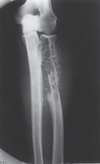
Fibrous Dysplasia.
Polyostotic fibrous dysplasia is seen in the radius in this child. Parts of this lesion have a hazy, ground-glass appearance, whereas others are more lytic appearing. A hazy, ground-glass appearance is often present in fibrous dysplasia, but just as often, the appearance can be purely lytic or even sclerotic.

Adamantinoma.
This mixed lytic and sclerotic process in the midshaft of the tibia is characteristic of fibrous dysplasia.
An adamantinoma has an identical appearance and should be considered in any tibial lesion that resembles fibrous dysplasia. Biopsy showed this to be an adamantinoma.

Enchondroma.
A lytic lesion in the phalanges is most commonly an enchondroma. This is the only location in the skeleton where an enchondroma does not contain calcified chondroid matrix.
These most often present with pathologic fractures, as in this example.

Bone Infarct.
These lytic lesions in the distal femurs with calcified, serpiginous borders are typical of bone infarcts. Occasionally, differentiating between a bone infarct and an enchondroma can be difficult on radiographs; however, in this example, infarcts are easily diagnosed.

Enchondroma.
This lesion in the distal right femur shows the stippled punctate calcification typical of chondroid matrix seen in an enchondroma.

Ollier Disease.
Multiple enchondromas are present throughout the hand. This is a typical example of Ollier disease.

Maffucci Syndrome.
Multiple enchondromas in the phalanges are associated with soft
tissue phleboliths. This combination of findings invariably represents hemangiomas and enchondromas in Maffucci syndrome.

Eosinophilic Granuloma (EG).
A well-defined lytic lesion is seen involving the mid- femur in this 20-year-old patient. Biopsy showed this to be EG.

Eosinophilic Granuloma (EG).
Well-defined lytic lesions are present throughout the pelvis in this 24-year-old patient. In addition to the lesion around the right hip, a lesion is seen at the right sacroiliac joint. Biopsy showed this to be EG

Eosinophilic Granuloma (EG).
This well-defined lytic lesion contains a bony sequestrum (arrow), which is typical of osteomyelitis or EG. Biopsy revealed this to be EG

Giant Cell Tumor.
A well-defined lytic lesion without a sclerotic margin is seen abutting the articular surface of the distal femur in a patient who has closed epiphyses. These are all characteristics of a giant cell tumor

Giant Cell Tumor.
This well-defined lytic lesion that does not have a sclerotic margin completely involves the greater trochanter. The apophyses have the same differential diagnosis as lesions in the epiphyses, which makes giant cell tumor a strong possibility in this example. Biopsy showed this to be a giant cell tumor.

Giant Cell Tumor.
A large, well-defined lytic lesion in the iliac wing is seen, which does contain a sclerotic margin and does not appear to abut any articular surface. The pelvis is a good location for giant cell tumor, which this proved to be at biopsy. The usual rules for giant cell tumors such as the presence of a nonsclerotic margin do not apply in flat bones.

Fibrous Cortical Defect.
A well-defined lytic lesion is seen in the medial metaphysis of this tibia (arrows), which is typical of a fibrous cortical defect.

Nonossifying Fibroma (NOF).
A large, well-defined lytic lesion, which is slightly expansile with scalloped sclerotic margins, is seen in the distal tibia in this young patient. This is a characteristic appearance of an NOF. The examination was obtained for a sprained ankle and not for
this asymptomatic lesion.

Nonossifying Fibroma (NOF).
A well-defined, expansile lytic lesion in the distal fibula is noted in this asymptomatic patient, which is characteristic of an NOF

Nonossifying Fibroma.
A: A well-defined, lytic lesion that is minimally expansile is seen in the distal tibia in this child who was examined for a sprained ankle.
B: A CT examination showed apparent cortical destruction (arrow), which was believed to be suggestive of an aggressive lesion.Biopsy showed this to be a nonossifying fibroma. Both CT and MR will often show apparent cortical destruction, which is merely cortical replacement by benign fibrous tissue.

Healing Nonossifying Fibroma (NOF).
A predominantly sclerotic lesion, which is minimally expansile and well defined, is seen in the proximal humerus in this child who is asymptomatic. This is a typical appearance of a disappearing or healing NOF. With time, this lesion will melt into the
normal bone and essentially disappear.

Nonossifying Fibroma (NOF).
This large, well-defined lytic lesion with faint sclerotic margins is seen in the distal femur. Because of its size, many thought it was not an NOF. The lesion underwent biopsy and was found to be an NOF

Osteoblastoma.
A lytic expansile lesion involving the right T-12 pedicle ( arrow) and transverse process is seen on this anteroposterior radiograph in (A) which is seen on the CT scan (B) to extend into the vertebral body. It has intact cortices and contains some calcified matrix. This is a classic example of an osteoblastoma of the spine

Metastatic Disease.
A well-defined lytic lesion is seen in the proximal femur in this 50-year-old patient who has pain associated with this lesion. Biopsy showed this to be a renal metastasis. A significant number of metastatic lesions can have a completely benign appearance, as in this example