BCS Flashcards

1
Q

What are the functions of calcium? (7)

A

Bone growth and remodelling Secretion - flux of intracellular calcium tells glands to release hormones Muscle contraction Blood clotting - calcium citrate used to help blood clot Co-enzyme Stabilization of membrane potentials - in heart and brain Second messenger/stimulus response coupling

2
Q

Where is calcium stored?

A

Mostly in the bone (99%) The rest is extracellular

3
Q

What regulates ionised ca2+

A

PTH and vitamin D

4
Q

What are the functions of phosphate? (4)

A

Element in: High energy compounds e.g. ATP, Second messengers e.g. cAMP Constituent of: DNA/RNA, phospholipid membranes. bone Intracellular anion Phosphorylation (activation) of enzymes

5
Q

Where is phosphate stored?

A

Skeleton 90% 9.97% intracellular (of which 50% is free, 50% is bound)

6
Q

How is free phosphate regulated?

A

Controlled by kidneys, PTH, FGF23

7
Q

What regulates the amount of calcium and phosphate in the blood?

A

The kidney Calcium - Distal CT Phosphate - Proximal CT

8
Q

Order of bone remodelling process (4). How long does the process take?

A

30 days Osteoclasts carving REABOSORPTION well Osteoblasts form osteod, to REVERSE the changes Osteoblasts initiate FORMATION of new bone Resting state

9
Q

What is an osteoclast?

A

Modified macrophage (from hematopoietic stem cellor mesenchymal stem cell)

10
Q

Review development of osteoclasts and osteoblasts

A

PICTURE

11
Q

Outline the hormonal control of bone remodelling

A

PTH activates osteoblasts, which controls bone reabsorption but releasing collagen and proteases IGF 1 is main actor

12
Q

How does the osteoblast stimulate differentiation of osteoclasts? What receptor is activated? What can inhibit differentiation?

A

Production of RANK ligand The osteoclast recursor has a RANK receptor that is activates via activation of nuclear kappa beta OPG binding inhibits differentiation

13
Q

Outline the role of osteoprotogenerin - what stimulates it?

A

also known as osteoclastogenesis inhibitory factor (OCIF) or tumour necrosis factor receptor superfamily member 11B (TNFRSF11B) False receptor to prevent activation of RANK receptor on osteoclast (to ultimately prevent bone reabsroption) Estrogen increases OPG expression (so dip in estrogen during menopause causes bone reabsoprtion)

14
Q

Describe the activities of bone as an endocrine organ

A

FGF23 acts in conjunction with PTH decrease phosphate reabsorption by down-regulating NaPi2a and NaPi2c expression in the brush border of the proximal tubule resulting in hyper-phosphaturia and hypophosphatemia regulation high phospate and high 1,25 di hydroxy vit D The primary mechanisms by which adiponectin enhance insulin sensitivity appears to be through increased fatty acid oxidation and inhibition of hepatic glucose production

15
Q

Outline the role of glucocorticoids, estrogen, calcitonin, thyroxine, vitamin A, androgens and GH on bone turnover

A
16
Q

Provide overview of parathyroid gland

A

4 glands on upper and lower poles of each lobe of the thyroid gland Supernumerary glands not uncommon (source of PTH excess?) 30-50 mg weight Chief cells and oxyphill cells Supplied by blood from the inferior thyroid arteries (thyroid surgery)

17
Q

What type of hormone is PTH? How can you measure it?

A

peptide (very short half life) Have to use sandwich assay to measure

18
Q

What happens to calcium levels in acidosis?

A

HIGH

19
Q

Outline the activity of the calcium sensing receptor? Where is it? What type of receptor? Ultimate results of activity

A
20
Q

If your calcium levels are two high, what should be happening to your PTH?

A

There shouldn’t be any in your system

21
Q

How does vitamin D regulate PTH production?

A

Calcitriol (1,25 OH2D3) inhibts replication and secretion of PTH To INCREASE calcium

22
Q

What are the direct actions of PTH on bone activity and in kidney (4)

A

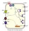
Stimulate osteoblasts to produce M-CSF and RANK ligand increased bone resorption Increase Ca2+ reabsorption in the distal convoluted tubule Increase phosphate excretion Increases 1-α hydroxylase in the proximal tubule

23
Q

REVIEW activity of PTH on kidney in more detail
A
24
Q

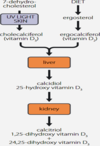
What is the active form of Vitamin D? How is it made?

A

D3 Cholesterol and UV light on skin / DIET
25
Q

What is the regulatory step in the production of vitamin D?

A

Hydroxylation on C1 in kidney So issue in kidney disease with calcium levels

26
Q

What are the actions of vitamin D in calcium homeostasis? (5)

A

Increases Ca2+ absorption in the gut Requires CaBP’s - synthesis stimulated by Vitamin D Synergises with PTH on bone Inhibits PTH synthesis Inhibits 1a-hydroxylase (feedback on itself)

27
Q

How is calcium absorbed through the gut? (2)

A

Paracellular transport – diffusion through tight junctions dependent on concentration gradient; does not require energy Transcellular – at apical region calcium enters cell through a selective calcium transporter (TRPV), binds to calbindin, transported across cell and extruded at the basolateral membrane by a sodium-calcium exchanger and a Ca2+/ ATPase transporter.

28
Q

What are the other roles of vitamin D (beyond gut, bone, kidney) (2)

A

PICTURE REGULATOR - possibly in cancer surveillance? Diabetes - making sure VitD levels okay to improve insulin resistance Vitamin D receptors found in more than 30 different cell types e.g keratinocytes in the skin, lymphocytes, macrophages, adipocytes, pancreatic β cells, cells of breast, testis, ovary, prostate, colon etc. Several tissues can also locally synthesis 1,25 (OH)2D from circulating 25(OH)D because they have 1α-hydroxylase (CYP27B1). Tissues/cells include macrophages and monocytes, keratinocytes, breat tissue, parathyroid, colon, placenta etc.

29
Q

Where is the FGF23 receptor expressed (3) What type of receptor is it?

A

Expressed pre- dominantly expressed in distal convoluted tubules epithelium of the choroid plexus in the brain parathyroid glands Klotho, a single-pass transmembrane protein

30
Q

What are the actions of FGF23 in calcium and phosphate homeostasis?

A

In kidney, down regulates phosphate channels in proximal CT which means phosphate gets excreted In kidney, it inhibits 1alpha-hydroxylase so decreases actions of vitamin D

31
Q

Major causes of hypercalcaemia (6)

A

1o Hyperparathyroidism Malignancy (PTHrP) Some other causes: Vitamin D related Excess intake Sarcoidosis, tuberculosis and other granulomatous diseases (10% cases extra renal conversion of 25-OH D  1,25-OH D) High bone turnover e.g. hyperthyroidism, immobilization Renal failure

32
Q

What are the symptoms of hypercalcemia? Neurologic, renal, MSK, CVS, GI

A
33
Q

Major causes of hypocalcaemia (6)

A

Vitamin D deficiency (2o hyperprathyroidism) Hypoparathyroidism (thyroid surgery) Chelation Pseudohypoparathyroidism - receptor defect Neonatal Activating mutation of Ca2+ receptor - FIHH

34
Q

Major causes of Vit D deficiency (5)

A

Liver/kidney disease (synthesis) Resistance to hormone (receptor) Mal-absorption Dietary insufficiency Poor exposure to sunlight Sun block Obesity Latitude Skin pigmentation (melanocytes)

35
Q

What are the typical symptoms / signs of Vit D deficiency (5)

A

Aches and pains in bones Proximal myopathy Mild hypocalcaemia - 2o hyperparathyroidism Hypophosphataemia and hyperchloraemic acidosis Bone deformities - osteomalacia

36
Q

Symptoms of hypocalcaemia

A
37
Q

Outline the endocrine response to LOW calcium

A
38
Q

What are the two types of bone? What are it’s two forms?

A

cortical bone (outside) and cancellous bone (inside) two forms: woven bone (the collagen fibres are randomly arranged, and this can be seen under polarized light. This is an immature form of bone that is produced when bone is formed rapidly eg in the neonate or in the early stages of fracture repair) and lamellar bone.

39
Q

Describe the structure of cortical bone

A

outer portion of long bones and vertebrae. Cortical bone is composed of long parallel columns, known as osteons, which are made up of concentric rings of bone, the lamellae, surrounding a central Haversian canal containing blood and lymphatic vessels.

40
Q

Describe the structure of cancellous bone

A

composed of a network of bony plates or struts called trabeculae. These connect with each other and to the endosteum of cortical bone. In adults, the spaces between the trabeculae are filled with either haemopoetic bone marrow or adipose tissue.

41
Q

What are osteocytes? What are their roles?

A

Osteocytes are terminally differentiated osteoblasts. These lie within lacunae and their cell processes connect with other osteocytes via cell processes that are in canaliculi. The function of osteocytes is thought to be mechanosensation.

42
Q

What are the three types of cartilage?

A

Hyaline cartilage is the most common type, and found in the nasal septum, larynx and tracheal rings, most articular surfaces of joints. Fibrocartilage is found in intervertebral discs and some joints such as pubic symphysis. Elastic cartilage occurs in the external ear, external auditory meatus, epiglottis, and parts of the laryngeal cartilage rings

43
Q

What muscles produce movements in the knee joint?

A

Extension: Produced by the quadriceps femoris, which inserts into the tibial tuberosity. Flexion: Produced by the hamstrings, gracilis, sartorius and popliteus. Lateral rotation: Produced by the biceps femoris. Medial rotation: Produced by five muscles; semimembranosus, semitendinosus, gracilis, sartorius and popliteus.

44
Q

What nerve is associated with foot drop?

A

peroneal nerve

45
Q

What is the role of the IT band? What about in knee stability?

A

The action of the ITB and its associated muscles is to extend, abduct, and laterally rotate the hip. Stabilises knee laterally

46
Q

ACL and PCL - what movements do these resist?

A

ACL prevents posterior movement, PCL prevents anterior movement

47
Q

Why is medial meniscus tear more common than lateral?

A

A medial meniscus tear is more common than a lateral meniscus tear, because it is firmly attached to the deep medial collateral ligament and the joint capsule. In addition, the medial meniscus absorbs up to 50% of the shock of the medial compartment, making the medial meniscus susceptible to injury.

48
Q

What are the 7 types of synovial joint? Example of each

A

Plane (gliding) – acromioclavicular Hinge (on one axis) – elbow (humero-ulnar joint) Pivot (around one axis) – atlanto-axial joint Bicondylar (mostly one axis, limited rotation) - knee Condylar (two axis at right angles) - wrist Saddle (two axis at right angles) – thumb (carpometacarpal) Ball & socket (multiple axis) – hip, shoulder

49
Q

What are the 3 fibrous joint types?

A

Sutures Gomphosis – teeth Syndesmosis – synarthroses – interosseous membrane

50
Q

What are the 2 cartilaginous joint types?

A

Symphysis (bones connected by cartilage) Synchondrosis (two layers of developing bone separated by cartilage)

51
Q

What is the function of the lateral collateral ligament?

A

stabilise the hinge motion of the knee, preventing excessive lateral movement

52
Q

What is the function of the medial collateral ligament?

A

stabilise the hinge motion of the knee, preventing excessive medial movement

53
Q

Which muscles are involved in extension of the leg?

A

Extension: Produced by the quadriceps femoris, which inserts into the tibial tuberosity

54
Q

Which muscles are involved in flexion of the leg?

A

Flexion: Produced by the hamstrings, gracilis, sartorius and popliteus

55
Q

Which muscles are involved in lateral rotation of the leg?

A

Lateral rotation: Produced by the biceps femoris.

56
Q

Which muscles are involved in medial rotation of the leg?

A

Medial rotation: Produced by five muscles; semimembranosus, semitendinosus, gracilis, sartorius and popliteus.

57
Q

In knee reflex test, which ligament is struck? Which nerve segments does it test?

A

Patellar ligament L2-L4

58
Q

What is the most common type of ankle sprains? Which ligaments are affected?

A

Inversion injuries anterior talofibular ligament (and all lateral ligaments)

59
Q

What are the structures within the popliteal fossa? (6)

A

Common fibular nerve Tibial nerve Popliteal artery Popliteal vein Sural nerve Lesser saphenous vein

60
Q

What are the different compartments of the leg?

A

posterior superficial (gastrocnemius and soleus) posterior deep (plantaris, popliteus, flexor digitorum longus, flexor hallucis longus, tibialis posterior) lateral (fibularis longus and brevis) Anterior (tibialis anterior, extensor digitorum longus, extensor hallucis longus,fibularis tertius)

61
Q

What are the motor roles of the deep and superficial peroneal nerves

A

Superficial fibular nerve: Innervates the muscles of the lateral compartment of the leg; fibularis longus and brevis. These muscles act to evert the foot.[6] Deep fibular nerve: Innervates the muscles of the anterior compartment of the leg; tibialis anterior, extensor digitorum longus and extensor hallucis longus. These muscles act to dorsiflex the foot, and extend the digits. It also innervates some intrinsic muscles of the foot.[6]

62
Q

What are the sensory roles of the deep and superficial peroneal nerves

A

Superficial fibular nerve: Innervates the skin of the anterolateral leg, and dorsum of the foot (except the skin between the first and second toes). Deep fibular nerve: Innervates the skin between the first and second toes

63
Q

What is the risk to the common peroneal nerve in injury?

A

The common peroneal nerve is in a particularly vulnerable position as it winds around the neck of the fibula.

64
Q

In what muscle does ‘shin splints’ occur?

A

Tibialis anterior

65
Q

What part of bones grow?

A

Epiphysis

66
Q

What is a grade 1 vs grade 2 vs grade 3 sprain?

A

Grade 1 - stretching, small tears

Grade 2 - Larger but incomplete tear

Grade 3 - complete tear

67
Q

What does a lipohaemosmosis indicate on imaging?

A

Diagnostic of fracture (just may not be visible) as means fat is leaking from bone

68
Q

When and whty are NSAIDs used?

A

First choice for MSK pain

Reduce pain, fever, inflammation so can be used for wide range of condition

Mostly work at reducing inflammation

69
Q

What factors drive the inflammation in MSK pain?

A

Lecocytes, cytokines, eicosanoids (arachidonic acid metabolites), others

70
Q

Descrobe overview of inflammatory process - which cells show up first? What are the ILs involved?

A

Short term response - Macrophages show up (release TNF and IL1) and call other cells to come

71
Q

REVIEW biochemical pathway of tissue injury. What are the different COXs?

A
72
Q

Outline the common adverse effects of NSAIDs and describe WHY these happen

A
73
Q

What are 2 cox-2 selective NSAIDs?

A

Celecoxib, meloxicam

74
Q

What are the issues with COX 2 selective NSAIDs?

A

Higher risk of myocardial infarction - so not safe long term

75
Q

What are some common NSAID interactions?

A
76
Q

How do you treat gout?

A

Aim - reduce pain and inflammation

NSAIDs - high dose diclofenac and naproxen

Colchicine - inhibits leukocyte micro-tubular formation / migration

Possibly steriofs

77
Q

What are the typical symptoms of rheumatoid arthritis?

A

painful, swollen

Increased temp

Stiff and painful in AM

Better after several hours

Symmetrical symptoms / pain

Systemic symptoms / other autoimmune conditions

78
Q

How do you treat rheumatoid arthritis?

A

Analgesia - paracetamol (with opoids if neded), NSAIDs

DMARDs - Methotrexate, sulphasalazine, azathioprine

Immunotherapy - anti-TNF (infliximab)

Surgery - pins in joints to stabilise them

79
Q

REVIEW normal range of motion in shoulder complex

A
80
Q

What are the main static stabilisers of the shoulder?

A

Gleno-humeral ligaments (superior, middle and inferior)

Others:

  1. Coraco-Humeral
  2. Ligament
  3. Glenoid Labrum
  4. GHJ bony anatomy
  5. Posterior capsule
  6. Negative intra-articular
  7. pressure
81
Q

What are dynamic stabilisers of the shoulder?

A
  • Rotator cuff
  • muscles
  • Long head of
  • Biceps
  • Deltoid
  • Scapulothoracic
  • musculature
82
Q

What is the most important stabilisier of the sternoclavicular joint?

A

Posterior capsular ligament

83
Q

What does the axillary nerve innervate? (2 examples)

A

Deltoid muscle
¡ Regimental Badge
sensation

84
Q

What does the musculocutaneous nerve innervate? (4 examples)

A
  • ¡ Short Head of Biceps
  • muscle
  • ¡ Brachialis muscle
  • ¡ Coracobrachialis muscle
  • ¡ Lateral Forearm sensation
85
Q

How do you investigate / manage a shoulder dislocation?

A
  • History & Examination
    • Neurovascular status
  • Reduce the joint if capable
  • Call for help
  • Sedation
  • Re-examine and x-ray after
  • reduction
  • Follow-up in specialist clinic
86
Q

What is the most common associated injury with dislocated shoulder?

A
  • Axillary nerve palsy
    • Axonal loss up to 48% in all dislocations*
    • Majority recover (12 - 40 weeks)
    • NCS/EMG at 6/52
      • If no change at 3/12 then ?Explore or Graft
    • Worse prognosis with increasing age
87
Q

Should someone have surgery after disclocating shoulder for first time?

A

IF they are under 30, have hada traumatic dislocation and no fracture, offerstabilisation within 6 weeks

88
Q

Describe impingement / bursistis - risk factors, symptoms, management

A
  • Middle age
    • Multi-factorial
    • § Rotator cuff tendonitis /
    • tendonosis
    • § Acromium bone spurs
    • § Capsular tightness
  • Symptoms
    • § Lateral upper arm pain
    • § Painful arc
    • § Night pain
    • § Functional pain
  • Management
    • Treat the cause
    • Rest / modify activity
    • Analgesia
    • Physiotherapy
    • Steriod injections
    • Surgery
89
Q
A
90
Q

Describe ACJ OA - risk factors, symptoms, management

A

Osteoarthritis of the acrmio-clavicular joint -

Primary Osteoarthritis

  • Post traumatic
    • § Weightlifters
    • § Over head workers
    • § All ages
    • § Male > females
  • Symptoms
    • § Pain directly over ACJ
    • § High arc pain
    • § Night pain
  • Management
  • Non-operative Treatment
    • § Rest
    • § Activity modification
    • § Analgesia
    • § Physiotherapy
    • § Steroid injections
  • Operative Treatment
    • § ACJ excision
91
Q

Describe rotator cuff tear - risk factors, symptoms, management

A
  • Traumatic vs Degenerative
    • § Over 40’s
    • § Overhead workers
    • § Inflammatory arthritis
  • Incidence
    • § 10% over 50
    • § 30% over 60
    • § 60% - 80% over 80
  • Symptoms
    • § Pain
    • ▪ Night
    • ▪ On movement
    • § Weakness on lifting / rotation
    • § Unable to lift arm
  • Investigations
    • § X-ray
    • § MRI vs US
  • Management
    • Non-operative
      • § Rest
      • § Activity modification
      • § Analgesia
      • § Physiotherapy
      • § Steroid injections
      • § Nerve blocks
        • ▪ Suprascapular
        • ▪ Lateral pectoral
    • Operative
      • Cuff repair
92
Q

Describe calcific tendonitis

A
  • Hydoxyapatite crystal deposition
  • 25% are bilaterally
  • Supraspinatus most common
  • Cause (they aren’t sure)
    • § Reactive condition
      • ▪ Trauma
    • § Endochondral calcification
      • ▪ Hypoxia
    • § Metaplasia of MSCs
      • ▪ Ectopic
  • Management
    • Non-operative
      • § Rest
      • § Activity modification
      • § Analgesia
      • § Physiotherapy
      • § Steroid injections
      • § Shock wave therapy
      • § Needle Lavage / Barbotage
      • ▪ US-guided
    • Operative
      • § Excision of calcium deposits
93
Q

Describe adhesive capsulitis - risk factors, associated diseases, management

A
  • Middle age
  • Disease of joint capsule
    • § Fibroblast proliferation
      • ▪ Transformation to myofibroblasts
      • ▪ Collagen deposition
  • 3 phases (Codman)
    • § Freezing
    • § Frozen
    • § Thawing
  • Associated with
    • § Diabetes mellitus
    • § Hypo/hyperthyroid
    • § Hyperlipidaemia
    • § Trauma
  • Management
    • Non-operative
      • § Rest (painful stage)
      • § Activity modification
      • § Analgesia
      • § Steroid injections
        • ▪ Glenohumeral joint
      • § Physiotherapy
        • ▪ once pain settling
      • § Hydrodilation
    • Surgery
      • § Manipulation
      • § Arthroscopic capsular release
94
Q

Describe osteoarthritis - risk factors, symtpoms, management

A
  • “Wear and Tear”
  • Degeneration of Synovial Joint
  • Reduced range of motion with pain
  • Symptoms
    • § Pain (Night)
    • § Grinding and clicking
    • § Stiffness
  • Management
    • Non-operative
      • § Rest on acute flair ups
        • ▪ Exclude inflammatory causes
      • ▪ Involve rheumatologist
      • § Activity modification
      • § Analgesia
    • Operative
      • § Arthroscopy
      • § Total Shoulder Replacement > Hemiarthroplasty
      • § Functioning Rotator Cuff required
95
Q
A