Basic ECG Interpretation Flashcards

1
Q

Describe a system for ECG interpretation

A

Rhythm and rate Cardiac axis PR interval QRS complexes ST segments and T waves QT interval

2
Q

Describe 2 methods for calculating rate on ECGs

A

300/no. of large squares between QRS complexes Count QRS complexes over strip (10s) and multiply by 6

3
Q

What is normal sinus rhythm?

A

1:1 ratio of P waves to QRS complexes

4
Q

What is respiratory sinus arrhythmia?

A

1:1 ratio of P:QRS but irregularity with respiration (faster with inspiration due to decreased vagal tone)

5
Q

Distinguish between atrial and ventricular ectopics in terms of their appearance on ECG

A

Atrial: early, narrow complex QRS followed by compensatory pause Ventricular: early, broad complex QRS

6
Q

Describe the ECG appearance of atrial fibrillation

A

Absence of P waves Irregularly irregular rhythm Always comment on ventricular response rate (>100 is rapid and

7
Q

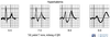
Describe the ECG appearance of atrial flutter

What is the ventricular response rate in atrial flutter and why? What does the ventricular response rate of atrial flutter suggest about the underlying cause?

A

“Saw tooth” p waves due to large re-entrant pathway in atrium

Length of the re-entry circuit corresponds to the size of the right atrium and atrial rate is therefore usually a regular 300 bpm (ventricular rate depends on the AV conduction ratio; commonly it is 2:1 resulting in a ventricular rate of 150 bpm)

Higher degree AV blocks can occur with atrial flutter, more commonly where the underlying cause is medications or underlying heart disease

Atrial flutter with 1:1 conduction can occur due to sympathetic stimulation or in the presence of an accessory pathway — especially if AV-nodal blocking agents are administered to a patient with WPW; this is associated with severe haemodynamic instability and progression to ventricular fibrillation

NB. The term “AV block” in the context of atrial flutter is something of a misnomer; AV block is a physiological response to rapid atrial rates and implies a normally functioning AV node

8
Q

Distinguish between narrow complex and broad complex tachycardias

A

Narrow: QRS 120ms

9
Q

When might an ECG be abnormal?

A

Cardiac pathology: conduction abnormalities, structural heart disease, ischaemia Systemic pathology: sepsis, PE, intracranial pathology, electrolyte disturbance)

10
Q

What leads are included in a standard ECG?

A

6 praecordial 6 limb (3 of which are augmented/derived)

11
Q

List 4 common cardiac presentations where an ECG would be an appropriate 1st line test

A

Chest pain Dyspnoea/HF Palpitations Syncope

12
Q

What additional leads can be added to a standard 12-lead ECG and what is the clinical scenario in which these may be indicated?

A

Right ventricular leads V4R-V6R (if suspecting right ventricular infarction Posterior leads V7-V9 (if suspecting posterior ischaemia)

13
Q

P wave

A

Atrial depolarisation

14
Q

QRS complex

A

Ventricular depolarisation (masks atrial repolarisation)

15
Q

T wave

A

Ventricular repolarisation

16
Q

What does the PQ interval represent and what is its normal length?

A

AV conduction time (measured from start of p wave to start of QRS)

17
Q

Normal equivalent time of a small square (1mm)

A

0.04s

18
Q

Normal equivalent time of a large square (5mm)

A

0.2s

19
Q

Normal speed of ECG

A

25 mm/sec (5 big squares = 1 sec)

20
Q

Normal duration and appearance of QRS complex

A

Usually not >0.1 sec in duration R waves are deflected positively and the Q and S waves are negative

21
Q

Where are p waves best seen?

A

Leads II, V1

22
Q

What changes are seen in left axis deviation?

A

Ladies Adore Diamonds (lead i ^, lead II/aVF down)

23
Q

What changes are seen in right axis deviation?

A

Rover Adores Digging (bone shape: lead I down, lead II/aVF ^)

24
Q

What does a normal cardiac axis look like?

A

Lead I ^` Lead II/aVF down

25
List 6 causes of left axis deviation (\>-30 degrees)
Left anterior hemiblock IHD Cardiomyopathy HTN WPW syndrome with R sided accessory pathway
26
List 5 causes of right axis deviation (\>+90 degrees)
Normal finding in children and tall thin adults RV volume/pressure overload (RV hypertrophy, ASD, VSD, PE) Lung pathology (COPD, PE) Dextrocardia (apex on R side of chest) WPW syndrome with L sided accessory pathway
27
List 5 causes of extreme R axis deviation
Lead transposition VT Emphysema Hyperkalaemia Paced rhythm
28
What is the normal duration of a p wave?
Duration
29
What is p pulmonale and what is it indicative of?
Increased p wave voltages Indicative of R atrial dilation
30
What is p mitrale and what is it indicative of?
Bifid p wave Indicative of L atrial dilation (later depolarisation of LA leads to 2 peaks in p wave)
31
What atrium depolarises first?
Right
32
In what demographic is 1st degree AV block common?
Normal individuals, especially young athletic people
33
What is the difference between 2:1 AV conduction block and complete heart block?
2:1 AV conduction block: every 2nd p wave is non-conducted Complete heart block: AV dissociation
34
What is the "key ECG concept"?
QRS complex is predominantly positive if vector of depolarisation moves towards that lead (and vice versa)
35
List 3 possible abnormalities of the QRS complex
Voltages (e.g. increased in LVH, decreased in cardiac amyloidosis) Q waves Conduction (e.g. L or R bundle branch blocks)
36
Identifying LVH on ECG
Sum of height of S in V1/V2 and R in V5 or V6 (whichever is larger) ≥35mm/3.5mV (7 large squares) Or R in aVL ≥11mm
37
What findings can be seen on ECG in R ventricular hypertrophy?
Right axis deviation R wave predominant in lead V1 (normally S wave dominant) Inverted T waves in right praecordial leads (V2, V3 in severe cases) Deep S in V6 Peaked P waves may also occur due to right atrial hypertrophy
38
When are Q waves normal?
Small septal Q waves in some LV leads may be normal (due to early depolarisation of septum from L to R) Q wave in lead III normal
39
What do pathological Q waves indicate?
Marker of electrical silence which implies established full thickness death of myocardium (i.e. scar) ≥25% height of the corresponding R wave (and/or \>40ms width and \>2mm in depth) Present in \>1 contiguous (adjacent) lead
40
Classic morphology for LBBB
QRS complex duration \>0.12ms (3 small squares) "WilliaM" (W in V1, M in V6) Inverted T waves lateral leads V5-V6, I, aVL No septal Q waves
41
Classic morphology for RBBB
QRS complex duration \>0.12ms (3 small squares) "MarroW" (M rSR' in V1, W in V6) Inverted T waves V2-V3 Slurred S wave in V6
42
Inferior leads
II, III, aVF
43
Anteroseptal leads
V1-V4
44
Lateral leads
V5-V6
45
When can ST segment changes NOT be interpreted?
If resting pre-existing LBBB
46
4 causes of ST segment changes
Myocardial ischaemia/infarction Pericarditis LVH with "strain" pattern Drugs e.g. digoxin toxicity
47
ECG changes in pericarditis
Widespread ST segment elevation
48
ECG changes in LVH with "strain" pattern
Increased voltages of QRS complexes Strain pattern: ST depression with T wave inversion in lateral leads V5-V6, I, aVL
49
Which coronary artery provides the posterior descending artery?
RCA and/or circumflex
50
High lateral leads
I, aVL
51
ECG signs of previous infarction
Q waves form if full thickness infarction T wave inversion often persists long term
52
Progression of ECG changes in AMI
Normal Peaked T wave (timing??) Increasing ST segment elevation (timing??) Q wave formation and loss of R wave T wave inversion
53
What aspects of the ECG are important to assess in a suspected STEMI?
Degree of ST elevation (measure number of mm - what is significant??) Assess coronary territory
54
Can ST depression be localised to a territory?
No (but elevation can be)
55
What do anteroseptal Q waves suggest? What is the possible significance of these?
Full thickness anteroseptal MI May be late presentation STEMI or old infarction with scar formation
56
What kind of ST depression is more concerning?
Horizontal or downsloping (suggests significant myocardial ischaemia)
57
ECG changes in digoxin toxicity
AF (irregular, no p waves) "Reverse tick" ST depression and T wave inversion in lateral leads
58
In which leads are T waves normally inverted? What does it represent and what is their typical appearance?
Leads V1 (sometimes V2), III, aVR Indicates ventricular repolarisation Usually rounded and asymmetrical
59
Causes of T wave abnormalities
Same as for ST segment changes (myocardial ischaemia/infarction, LVH with "strain" pattern, digoxin toxicity) Systemic issues (e.g. electrolyte imbalances - K+/Mg2+/Ca2+)
60
What changes to the T wave suggest ischaemic changes? Which are most useful in diagnosing myocardial ischaemia?
Tall T wave Biphasic T wave (useful) Inverted T wave (useful) Flat T wave
61
ECG changes in hypokalaemia
Flattening of T waves U waves
62
ECG changes in hyperkalaemia
Tall, peaked T waves Widening of QRS
63
How is the QT interval measured? What does it represent? In which leads is the T wave optimised? What is its usual length?
Measured from start of Q to end of T Represents ventricular depolarisation and repolarisation (Na+ influx and K+ efflux) Leads V3, V4 or II optimise T wave QT interval usually less than half the R-R
64
How is the corrected QT interval calculated?
QT(corrected) = QT/(square root of R-R)
65
What is the normal QT(corrected) for women and men?
Women:
66
What is the concern with a prolonged QT interval?
Predisposes to torsades de pointe
67
List some acquired causes of prolonged QT
MED: MI Electrolyte imbalance Drugs
68
What drugs can predispose to prolonged QT?
Sotalol Amiodarone Azithromycin Amitriptyline Clozapine
69
What abnormalities are seen with inherited prolonged QT?
Defective sodium or potassium channels
70
What is Wolff-Parkinson White?
An accessory pathway (macro-reentrant circuit) which bypasses the AV node, leading to earlier (pre-) excitation of the ventricle ("delta wave" with short P-R interval) than would occur with normal conduction May lead to rapid regular tachycardias
71
ECG changes in WPW
Short P-R interval "Delta" wave (pre-excitation of ventricle)
72
What is the abnormality? What underlying pathology is this consistent with?
Early, narrow QRS followed by compensatory pause Consistent with atrial ectopics (premature atrial complexes)
73
Causes of narrow complex vs broad complex tachycardia??
??
74
What is the abnormality? What underlying pathology is this consistent with?
Irregularly irregular rhythm, absence of p waves, rapid ventricular response rate (\>100 bpm) Consistent with AF
75
What is the abnormality? What underlying pathology is this consistent with?
"Saw tooth" appearance of p waves due to large re-entrant pathway in atrium, length of the re-entry circuit corresponds to the size of the right atrium and atrial rate is therefore usually a regular 300 bpm (ventricular rate depends on the AV conduction ratio; commonly it is 2:1 resulting in a ventricular rate of 150 bpm) Consistent with atrial flutter
76
What is the abnormality? What underlying pathology is this consistent with?
AF (irregular, no p waves) with "reverse tick" ST segment depression and T wave inversion in lateral leads Consistent with digoxin toxicity
77
What is the abnormality? What underlying pathology is this consistent with?
QRS complex duration \>0.12s ,"WilliaM" (W in V1 but not always obvious, and M in V6), inverted T waves in lateral leads V5-V6, I, aVL; no septal Q waves Consistent with LBBB
78
What is the abnormality? What underlying pathology is this consistent with?
Increased voltage of QRS complexes; strain pattern - ST depression with T wave inversion in lateral leads V5-V6, I, aVL Consistent with LVH with "strain" pattern
79
What is the abnormality? What underlying pathology is this consistent with?
Horizontal ST depression Consistent with myocardial ischaemia
80
What is the abnormality? What underlying pathology is this consistent with?
Widespread saddle-shaped ST elevation Consistent with pericarditis
81
What is the abnormality? What underlying pathology is this consistent with?
QRS complex duration \>0.12s, "MarroW" (M - rSR' - pattern in V1 and W in V6), inverted T waves lead V2-V3, slurred S wave in V6 Consistent with RBBB
82
What is the abnormality? What underlying pathology is this consistent with?
Right axis deviation; R wave predominant in lead V1 (normally S wave dominant); inverted T waves in right praecordial leads V2, V3 (severe cases); deep S in V6; peaked p waves may also occur due to right atrial hypertrophy Consistent with RVH
83
What is the abnormality? What underlying pathology is this consistent with?
Early, broad complex QRS Consistent with ventricular ectopics
84
What is the abnormality? What underlying pathology is this consistent with?
Short PR interval; delta wave (reflects pre-excitation of the ventricles) Consistent with Wolff-Parkinson White syndrome
85
Causes of atrial and ventricular ectopics
Both are normal electrophysiological phenomenon usually not requiring Ix or treatment (but which may trigger a re-entrant tachydysrhythmia in patients with an underlying predisposition); frequent ectopics may cause palpitations and a sense of the heart "skipping a beat" Both may be caused by sympathomimetics (anxiety, caffeine, B-agonists), hypokalaemia, hypomagnesaemia, digoxin toxicity, myocardial ischaemia *NB Frequent ventricular ectopics are usually benign EXCEPT in the context of prolonged QT where they may predispose to Torsades de Pointes by causing an "R on T" phenomenon*
86
Anatomy of the myocardial conduction system