Approach to Eye Complaint Flashcards
What are the possible associated sx of eye complaint?
- pain
- drainage
- itching/burning
- vision change
- blurry vision
- flashing lights
(bolded terms are emergent)
What are the conditions of PMHx that would be relevant within an eye complaint?
- glaucoma
- diabetes
- thyroid dz
- ASCD
- collagen vascular dz
- HIV
- IBD
What external areas of the eye should you inspect during the exam?
- eyebrows
- periorbital area
- lashes
- lacrimal apparatus
- conjunctiva
- cornea
What should you look for when inspecting eyebrows?
- symmetry
- plucking or falling out
- scaly skin (seborrheic dermatits)
- scars
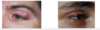
What should you look for when inspecting periorbital area?
- edema, bruising, injury
- allergic shiners (top photo)
- xanthelasma (cholesterol level, 2nd photo)
- proptosis/exophthalmos (bulging eyes, usually a/w hyperthyroidism, 3rd photo)
- dacrocytosis
- rash (to hairline consider shingles, pustules consider acne or insect bites; bottom photo)

What should you look for when inspecting eyelids/eyelashes?
- incomplete eyelid closure (possibly proptosis (top photo) or perioribital edema)
- flakiness/crustiness (blepharitis)
- erythema (contact derm, allergies, ifxn)
- swelling
- scabs/new lesions (BCC (2nd photo) or SCC)
- eyelid inversion (entropion, 3rd photo) or eversion (ectropion, last photo), ectropion seen in elderly, prior surgeries, previous ifxns, or genetic disorders
- ptosis
- chalazion or hordeolum
(palpation of eyelid may reveal chalazion, hard sensation/pain may represent hyperthyroidism, glaucoma, or retrobulbar tumor)

What are the different underlying causes of ptosis?
(ptosis: drooping of upper eyelid due to muscle abnormality)
- congenital: absent levator (won’t be tested on other causes)
- mechanical: inflammation, eyelid tumors, dermoid cysts
- aponeurotic: dehiscence of aponeurosis connecting levator muscle to eyelid
- neurologic: CN III palsy (ptosis, diplopia, ophthalmoplegia, down and out gaze), Horner’s syndrome (won’t be tested on this), other causes (botulinum toxin or myasthenia gravis)
- myogenic: won’t be tested on causes
What is the difference between a chalazion and a hordeolum (stye)?
- chalazion: blocked meibomian gland, usually nontender unless inflamed, in the lid, not an infection (left photo)
- hordeolum (stye): bacterial infection of meibomian gland, tender/painful, along the lashline (right photo)
What are the possible causes, symptoms, and tx for blepharitis?
- causes: bacterial (s. aureus) most common, inflammatory skin conditions (psoriasis, seborrheic derm, rosacea, eczema), or allergens (cosmetics, contacts)
- symptoms: red swollen itchy eyes, gritty/burning sensation, excessive tearing, blurred vision improves w/ blinking, flaking/scaling eyelids (in photo)
- treatment: warm compress, eyelid washing w/ dilute baby shampoo, artifical tears, topical abx (if patient doesn’t respond to other tx)

What are the possible conditions a/w lacrimal apparatus?
- clogged tear duct: transient, common in infants (most outgrow), keep eye clean and warm compresses
- dacrostenosis: narrowing of nasolacrimal duct, “milk” the duct for tx, may require probing duct
- dacrocystitis (photo): infection of lacrimal duct, common in newborns and elders, requires systemic abx and occasional probing duct

What are the 3 common abnormal findings of conjunctiva?
- erythema (subconjunctival hemorrhage)
- purulence (pink eye, conjunctivitis)
- pterygium (tissue growth)
(normal conjunctiva should be clear)

What are the 3 possible causes of conjunctivitis?
- allergic: mild, bilat sx; gritty, pruritic, irritated eyes w/ clear discharge
- viral: mild-mod bilat sx; gritty, burning, irritated eyes w/ clear discharge; eyes matted shut in morn
- bacterial (photo): usually unit w/ copious amnts of purulent drainage throughout entire day; drainage reaccumulates minutes after cleaning
(tx not included b/c we will not be tested on this)

What should you look for when inspecting cornea?
(test corneal sensitivity (CN V sensory and CN VII motor) w/ cotton ball)
- cornea should be clear
- brown tint: blood from trauma in anterior chamber
- white scar: previous abrasion/ulcer
- corneal abrasion
- white line encircling iris (arcus senilis)
How do you look for a corneal abrasion and what are possible causes?
- visualization via fluorescein stain and blue light (look for/remove foreign body)
- possible causes: foreign body or herpes simplex keratitis (pathognomonic dendritic lesion, leading cause of blindness worldwide)

- whitish linear (lipid deposition) encircling the colored iris
- common over 60 y/o
- if < 40 y/o, check cholesterol levels
arcus senilis

How should the lens appear with the red light reflex?
- lens should be clear, red color is from the retina
- yellow or gray lens: possibly cataract, can be nml in persons w/ increased melanin, but should be symmetric
- brown speckles: possibly cataract

- sudden increase in intraocular pressure, considered a medical emergency
- underlying cause of increased pressure: failure of aqueous flow from ciliary body into irido-corneal junction
- acute, severe pain; decreased vision
- pupil will be dilated and fixed
- tx: ophthalmologist referral
acute angle closure glaucoma

How should the sclera appear on exam?
- normal sclera is white
- brown: can be birthmarks (increased melanin), can be a/w increased risk for glaucoma
- blue: inherited, brittle bone dz
- yellow: icterus, neonatal, liver dz, pancreatic cancer, GB dz
What is the purpose of the eye cover/uncover test?
- have patient cover 1 eye while staring straight at a fixed point
- movement in uncovered eye means tropia is present (eye moves opposite direction of tropia, either esotropia (eye turned in) or exotropia (eye turned out))

How do you treat esotropia/exotropia (aka lazy eyes) and what can happen if left untreated?
- tx: eye patching over unaffected eye in young children, if this fails then surgery
- if not treated: brain chooses to focus w/ unaffected eye and affected eyes loses vision (amblyopia)
How do you use an ophthalmoscope?
- use the same eye and hand as the eye of the patient you are examining
- have patient look at fixed point behind your shoulder
- start 15 inches away from patient and move closer until you are almost cheek to cheek
What will you see in a normal ophthalmoscopic exam?
- veins and arteries (veins more defined/darker/larger, arteries brighter/smaller)
- optic disc (yellowish orange oval, color can vary w/ ethnicity)
- macula (darkened area)
- margins should be sharp

- increased intracranial pressure that results in intra-axonal edema along optic nerve, leading to swelling and engorgement of optic disc
- possible causes: intracranial hemorrhage, meningitis, trauma, mass lesion
papilledema

- increased intraocular pressure within eye leads to backward depression of the disc and atrophy
- base of the backward depression of disc will be pale in appearance
- normal cup to disc ratio is 0.4, ratio of 0.7 suggests glaucoma
glaucomatous cupping


