Appendix 4 Flashcards
(23 cards)

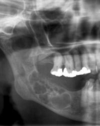
Dentigerous cyst description; unilocular radiolucency- pericoronal
etiology; cyst that develops by the separation of the follicle from around the crown of an unerupted tooth, develops by fluid accumulation between reduced enamel epithelium (REE) and the tooth crown
Tx; enucleation of cyst along with unerupted tooth
other; Most common developmental cyst, most often involves mandibular 3rd molars
VS Hyperplastic Dental Follicle- upon biopsy there is no cyst
(normal dental folcile- no tx-1-3mm)
(3-5mm could be a dentigeours cyst- biopsy to find out)
(5 or above mm its a dentigerous cyst)

Periapical Granuloma; unilocular radiolucency-periapical location
Description; radiographic pattern- Radiolucent lesion, Variable size, Symmetrical, Well defined, Loss of lamina dura, root resorption can be seen.
clinically- Most are asymptomatic, insensitive to percussion, no response to thermal or electric pulp test
etiology; prostaglandins activate osteoclasts to resorb surrounding bone, leading to periapical radiolucency. With time chronic inflammatory cells begin to dominate. granulation tissue- dense lymphocytic infiltrate (blue) that is intermixed with plasma cells (clock face nuclei).
tx; RCT, or extractaction followed by curettage of all apical soft tissue.
other;the most common periapical pathosis

Periapical cyst-Unilocular Radiolucencies: Periapical Location
Description; radiographic pattern- identical to that of a periapical granuloma, Radiolucent lesion, Variable size, Symmetrical, Well defined, Loss of lamina dura, root resorption can be seen. Significant growth is possible, May show static behavior or very slow growth, can be residual upon affected tooth removal
clinically- Most are asymptomatic, insensitive to percussion, no response to thermal or electric pulp test
histology- spiderweb epithelium
etiology; Inflammatory stimulation of epithelium in the area (Rests of Malassez)
tx; RCT, or extractaction followed by curettage of all apical soft tissue.

Antral Pseudocyst
Common finding on panoramic radiographs on 2-15% of population.
Mostly asymptomatic.
Increased prevalence during winter months.
Appears as dome-shaped, faintly radiopaque lesion arising from floor of maxillary sinus.
Develops due to accumulation of serum exudate (not mucous) beneath maxillary sinus mucosa, causing sessile elevation.
No treatment necessary.

Condensing Osteitis/Focal Sclerosing Osteomyelitis
Localized area of bone sclerosis associated with apices of teeth with pulpitis.
Association with inflammation distinguishes it from idiopathic osteosclerosis diagnosis.
Increased RO adjacent to tooth apex that has a thickened PDL.
No RL border distinguishes it from cemento-osseous dysplasia.
No clinical expansion of bone.
85% regress after odontogenic infection is eliminated.
Associated with teeth that have had extensive treatment i.e. using condenser.

Idiopathic Osteosclerosis
Etiology: Focal area of increased radiodensity that is of unknown cause and cannot be attributed to anything else
Tx: - no tx indicated. Only biopsy if there are symptoms, continued growth or cortical expansion

Odontoma-Complex
Most common odontogenic tumor, but once removed it won’t come back.
Complex; Conglomerate mass of enamel and dentin that bears no resemblance to a tooth.
Asymptomatic, usually discovered on radiograph taken to diagnose failure of a tooth to erupt.
Average age is 15.
Considered developmental anomalies (hamartomas) not true neoplasms.
Tx is simple local excision; once removed it won’t come back.

Odontoma-Compound
Most common odontogenic tumor, but once removed it won’t come back.
Compound; Composed of multiple tooth like structures.
Asymptomatic, usually discovered on radiograph taken to diagnose failure of a tooth to erupt.
Average age is 15.
Considered developmental anomalies (hamartomas) not true neoplasms.
Tx is simple local excision; once removed it won’t come back.

Tonsilloliths
Pharyngeal tonsillar crypts which are filled with desquamated keratin and foreign material.
Usually discovered as ROs in the midportion of the ascending ramus.
Secondarily become colonized with bacteria, calcify and develop foul smell.
Can promote recurrent tonsillar infections.
Usually asymptomatic.
Tx:
At home: Gargle warm salt water and/or use water jet. Bathroom surgery.
In office: Enucleation, local excision, or tonsillectomy is definitive.

Torus/Exostoses
Etiology: localized bony protuberance arising from cortical plate
- Best Known: Torus Palatinus, Torus Mandibularis
- Other Types: Buccal Exostoses, Palatal Exostoses, Solitary Exostoses
Tx: No tx except removal if trauma is an issue

Traumatic Bone Cyst
Description: “Scalloping.” Not true “cyst” - no epithelial lining.
Etiology: Trauma-Hemorrhage Theory is most widely accepted theory.
- Trauma to the bone which is insufficient to cause a fracture results in intraosseous hematoma
- If the hematoma does not undergo organization & repair, it may liquefy and result in a defect
Tx: surgical exploration and curettage
Provide a Differential Diagnosis

CEOT/AFO/COC/AOT
DDx: Mixed RL/RO
CEOT - Calcifying Epithelial Odontogenic Tumor
Description:
AKA Pindborg Tumor
“Driven-Snow Pattern”
Avg age 40
Md>Mx, F = M, Post > Ant
Liesegang rings (amyloid-like areas), Positive for Congo Red Test
Etiology: Tumor of Odontogenic Epithelium
Tx: Conservative Local resection with a narrow rim of bone or curettage
COC - Calcifying Odontogenic Cyst
Description:
AKA Gorlin Cyst
Avg Age 35
Etiology: Unknown. Classified by WHO as odontogenic tumor but listed under developmental odontogenic cyst in book
Tx: Simple enucleation or simple surgical excision
AOT - Adenomatoid Odontogenic Tumor
Description:
“Snowflake Calcifications”
Avg age 10 - 20 (uncommon over 30)
Mx>Md, F>M (2:1)
Etiology: Tumor of odontogenic epithelium
Tx: Enucleation
AFO - Ameloblastic Fibro-Odontoma
Description:
Avg age 10
Post Jaw
Etiology: Mixed Odontogenic Tumor (odontogenic epithelium + odontogenic ectomesenchyme)
Tx: Conservative Curettage

Stafne defect
aka Lingual Mandibular Salivary Gland Depression
Focal concavity of the cortical bone on the lingual surface of the mandible caused by a portion of the submandibular salivary gland
Treatment-None
What is this?

Ameloblastoma
Most common clinically significant odontogenic tumor
85% occur in mandible molar-ascending ramus region (except desmoplastic which occurs in anterior maxilla)
Multilocular RL
Buccal-Lingual cortical expansion
Histopathologic features-Palisading basal layer, Reverse polarity
Treatment
Varies from enucleation and curettage to en bloc resection
Marginal resection is the most widely used treatment-1.5 cm beyond what is visible radiographically
What is this?

Central Giant Cell Granuloma
Non-neoplastic lesion
**More common in the anterior jaw; frequently cross the midline**
Cherubism
Treatment is curettage with a recurrence of 20%
What is this?

Focal Cemento-Osseous Dysplasia
90% females
Average age = 40
More common in Caucasians
What is this?

Periapical Cemento-Osseous Dysplasia
90% female
70% African American
Periapical region of anterior mandible
PDL will be intact, lesion will not fuse to roots
What is this?
Florid Cemento-Osseous Dysplasia
90% Female
90% African American
Tendency to be bilateral and symmetrical
What is the treatment for cemento-osseous dysplasia?
For Florid and Periapical, diagnosis can be made from distinctive clinical and radiographic features-No biopsy required
Focal may require surgical investigation because features are less specific
When lesions are in RL phase they usually don’t cause any problems
Once in the RO phase lesions are hypovascular and prone to necrosis and secondary infection-frequent recall
Identify etiology/TX/other important info.

Nasoplatine Duct Cyst
ETIOLOGY
-Two passageways persist in midline between the primary and secondary palates
IMPORTANT INFO
- MOST COMMON NON-ODONTOGENIC cyst
- Radiolucency in/near anterior maxilla between apical central incisors (NO RESORPTION)
TX
- NEVER sit and watch
- Biopsy is mandatory (cannot diagnose radiographically)
- Surgical enucleation
Identify etiology/TX/other important info.

Residual Cyst
ETIOLOGY
- Cyst that persist at the site of a previous tooth extraction.
- inflammation and infection that stimulate rests of malassez
IMPORTANT INFO
-As the cyst ages, cellular components degenerate and can lead to dystrophic calcification and central luminal radiolucency
TX
-Surgical Excision
Identify etiology/TX/other important info.

Odontogenic Keratocyst
ETIOLOGY
Arises from cell rests of the dental lamina
IMPORTANT INFO
30% recurrence rate (up to 10 yrs after surgery)
Unilocular (smaller lesions)/Multilocular (larger lesions)
CAN CROSS MIDLINE
Young patients (<20 yrs) with OKC, should be evaluated/questioned for GORLIN SYNDROME
HISTO!!!
Basal cell layer shows palisading and is hyperchromatic
Epithelial surface is 6-8 layer thick and is corrugated
TX
Enucleate and curettage
Identify etiology/TX/other important info.

Lateral Periodontal Cyst
ETIOLOGY
Arises from rests of the dental lamina (rest of Serres)
Developmental odontogenic cyst along lateral root surface
IMPORTANT INFO
Intrabony counterpart of the gingival cyst of an adult
MANDIBULAR PREMOLAR-CANINE-LATERAL INCISOR area
TX
Conservative enucleation
