Anterolateral abdominal wall Flashcards

1
Q

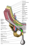
The abdominal cavity is larger than it appears externally. Why?

A

because the respiratory diaphragm arches superiorly under the rib cage.

2
Q

The abdominal cavity contains the……… cavity and………….. viscera.

A

peritoneal

abdominal

3
Q

What is Important to remember about the domes of the diaphragm?

A

The diaphragm has a right dome that rises as high as the 5th rib and a left dome that rises as high as the 5th intercostal space during expiration (So the RIGHT dome is HIGHER!)

4
Q

The upper abdominal organs (ex: liver and spleen) are protected by the lower ribs and costal cartilages of the thoracic wall. How may they be injured?

A

By fractures of the lower ribs

5
Q

The abdominal wall consists of………… vertebrae posteriorly, and the wings (alae) of the ilia laterally. There are layers of what located anterolaterally in the interval between the ribs and pelvis?

A

all 5 Lumbar vertebrae

skeletal muscle and aponeurosis

6
Q

For the localization of internal organs or patient symptoms, the abdomen is often subdivided into how many quadrants?

A

4

7
Q

For more precise localization, the abdomen may be subdivided into nine regions by two vertical midclavicular planes, and two horizontal planes:

A

The subcostal plane: through the 10th costal cartilages (or the trans pyloric plane) - The trans tubercular plane: through the tubercles of the iliac crest (level of Lv5)

8
Q

The superficial fasciaof the anterolateral abdominal wall Contains a variable amount of fat, and also contains cutaneous nerves, including branches of:

A

–Thoracoabdominal nerves (T7-11). This is why pain from the lower thoracic wall (ex: pleurisy) may be referred to the abdomen. Also note that T10 innervates the umbilicus level

–Subcostal nerve (T12)

–Iliohypogastricand ilioinguinal nerves (L1)

9
Q

Arteries of the anterolateral abdominal wall include which superficial vessels?

A
  • Superficial epigastric arteries
  • superficial circumflex iliac arteries (from the femoral artery)
10
Q

The deeper arteries of the abdominal wall include what?

A

– Inferior epigastric artery (external iliac) and superior epigastric artery (internal thoracic). Their anastomosis is a potential source of collateral circulation

–Deep circumflex iliac artery (from the external iliac artery)

* Remember that veins of similar names accompany the arteries.

11
Q

There are superficial collateral routes for venous return to the heart if the inferior or superior vena cava is obstructed. Desribe this!

A

(on each side) the anastomosis of thesuperficial epigastric vein (femoral vein) with the lateral thoracic vein (axillary vein) form the thoracoepigastric vein.

12
Q

When it comes to lymphatic drainage from the anterolateral abdominal wall, the superficial lymphatic vessels located………… drain mainly upward to axillary lymph nodes, and the ones located……………. drain downward to the superficial inguinal lymph nodes.

A

above the umbilicus

below the umbilicus

13
Q

Deep lymphatic vessels accompany what?

A

deep veins of the abdominal wall

14
Q

The superficial fascia (subcutaneous connective tissue) of the anterior abdominal wall consists of how many layers?

A
  • Above the level of the umbilicus it consists of a single fatty layer
  • Inferior to the umbilicus it consists of two layers
15
Q

The superficial fascia (subcutaneous connective tissue) of the anterior abdominal wall that is inferior to the umbilicus consists of two layers. What are these 2 layers?

A

– A superficial fatty layer, or Camper’s fascia

– A deep membranous layer, or Scarpa’s fascia

16
Q

Scarpa’s fascia in men is continuous with the superficial fascia of the scrotum and the perineum. Why is this important clinically?

A

extravasated urine from a ruptured penile urethra, or an infection, may spread upward into the anterior abdominal wall deep to the Scarpa’s fascia!

* however, fluid doesn’t spread downward into the thigh.

17
Q

There are 5 muscles in the anterolateral abdominal wall, ……….flat muscles whose fibers begin posterolaterally, pass anteriorly and are replaced by an aponeurosis towards the midline, and……… vertical muscles near the midline.

What is the nerve supply for these muscles?

A

3

2

•Nerve supply : anterior rami of T7-T12 spinal nerves

18
Q

The 3 flat muscles on each side of the anterolateral abdominal wall are:

A
  • external oblique
  • internal oblique
  • transversus abdominis

Nerve supply : anterior rami of T7-T12 spinal nerves

19
Q

The 2 vertical muscles near the abdominal midline are:

A

rectus abdominis and pyramidalis.

Nerve supply : anterior rami of T7-T12 spinal nerves

20
Q

The external oblique muscle arises from the lower 8 ribs and courses what direction?

It has posterior fibers inserting into the iliac crest and a broad EXTERNAL OBLIQUE APONEUROSIS anteriorly that helps form the anterior layer of the rectus sheath.

At the midline, linea alba aponeurotic fibers intersect with those of the other side.

A

inferomedially

21
Q

Between the anterior superior iliac spine and the pubic tubercle, the external oblique aponeurosis has a rolled-under inferior free margin. What does this form?

A

The inguinal ligament.

22
Q

The internal oblique muscle arises from thoracolumbar fascia, iliac crest, and the lateral 1/2 of the inguinal ligament. It has fibers coursing…………… at a right angle to the external oblique and continuing into the internal oblique aponeurosis.

It helps form the rectus sheath and intersects where?

A

superomedially

The linea alba

23
Q

The most inferior fibers of the internal oblique join those of the deeper………… muscle to form what?

A

transversus abdominis

the conjoint tendon (falx inguinalis, seen at red arrow in pic) arching over the spermatic cord (or the round ligament of the uterus) to attach to the pubic crest and pectin pubis.

24
Q

The transversus abdominis muscle Runs mainly transversely but with the lowest tendinous fibers arching downward to help form what tendon?

A

The conjoint tendon (red arrow)

25
Q

What muscle has three or more tendinous intersections (bulges of the intervening muscle) that make up “6-pack abs?”

A

The rectus abdominis muscle

26
Q

The rectus abdominis is enclosed within the connective tissue rectus sheath formed by the aponeuroses of which muscles?

A

the three flat abdominal muscles.

27
Q

The rectus abdominis is separated from the muscle of the other side by the midline called the “linea alba,” which is formed by the decussation of aponeurotic fibers of the obliques and transversus muscles. What is important to remember about the linea alba?

A

no major nerves or blood vessels cross the linea alba!

28
Q

The rectus abdominis is laterally bounded by a shallow convex groove called the linea……….., which is more obvious in thin or muscular individuals.

A

semilunaris

29
Q

The rectus sheath completely encloses the upper 3/4th of the rectus abdominis. It covers the………. surface of the lower 1/4th of the rectus abdominis, and the muscle at this point is in direct contact with the transversalis fascia.

A

anterior

30
Q

When it comes to formation of rectus sheath surrounding the upper 3/4th of the rectus abdominis, the……….. wall of the sheath consists of the aponeurosis of external oblique and half of the aponeurosis of internal oblique, which splits at the lateral margin of rectus abdominis .

The…………… wall of the rectus sheath consists of the other half of aponeurosis of internal oblique and the aponeurosis of transversus abdominis.

A

anterior

posterior

31
Q

When it comes to formation of rectus sheath surrounding the lower 1/4th of the rectus abdominis, the point of transition is marked by an arch of fibers called what?

A

The arcuate line.

32
Q

What are the Contents of the Rectus Sheath?

A
  1. Rectus abdominis
  2. Pyramidalis
  3. Superior epigastric artery & vein
  4. Inferior epigastric artery & vein
  5. Distal portions of anterior rami of T7-T12 spinal nerves
  6. Lymphatic vessels
33
Q

What is the second vertical muscle of the anterolateral abdominal wall, which is small and triangular shaped, and tenses the linea alba?

A

Pyramidalis

* It may or may not be present.

34
Q

Involuntary muscle spasms of the anterolateral abdominal wall muscles may occur due to inflammation that irritates their nerve supply (ex: in acute appendicitis) (rigidity)

Involuntary contraction may occur when?

A

during palpation with cold hands or voluntarily for protection (guarding).

35
Q

There is relaxation of the anterolateral abdominal wall muscles during…….. and contraction during…………..

A

inspiration

forced expiration

36
Q

The anterolateral abdominal wall muscles also increase intra-abdominal pressure, which is important for what processes?

A
  • Emptying the urinary bladder (micturition) and rectum (defecation)
  • Coughing and sneezing
  • Emesis (vomiting)
  • Giving birth (parturition)
37
Q

The anterolateral abdominal wall muscles are important for movement. Bilateral contraction results in what?

Muscles of one side contracting does what?

When one internal oblique and the contralateral external oblique contracting together, this produces rotation of the trunk to the side of the…………. (ex: moving the left shoulder toward the right hip).

A

flexion of the trunk

lateral flexion of the trunk to the ipsilateral side

internal oblique

38
Q

The inguinal region is an area of weakness within the inferior part of the anterolateral abdominal wall. It contains the inguinal canal, which transmits the large………… in males and the thin round……………. in females.

A

spermatic cord

ligament of the uterus

39
Q

The inguinal canal is an oblique passage through the abdominal wall. It extends inferomedially from the………………. (an outpouching of transversalis fascia) to the………………., a split in the external oblique aponeurosis.

A

deep inguinal ring

superficial inguinal ring

40
Q

The deep inguinal ring is located just above the inguinal ligament and immediately…………… to the inferior epigastric vessels.

A

lateral

41
Q

The superficial (external) inguinal ring is the end of the inguinal canal and is superior to the pubic tubercle. It is a triangular opening in the aponeurosis of the external oblique, with its apex pointing superolaterally and its base formed by the pubic crest. The two remaining sides of the triangle (called the……………..) are attached to the pubic symphysis and the pubic tubercle, respectively. At the apex of the triangle the two crura are held together by crossing (intercrural) fibers, which prevent further widening of the superficial ring.

A

the medial crus and the lateral crus

42
Q

When it comes to the boundaries of the inguinal canal, what forms the anterior and posterior wall?

A
  • Anterior wall: formed mainly by the external oblique aponeurosis with reinforcement laterally from the internal oblique aponeurosis.
  • Posterior wall: formed mainly by transversalis fascia reinforced medially by the conjoint tendon.
43
Q

When it comes to the boundaries of the inguinal canal, what forms the roof and the floor?

A
  • Roof: formed by the arching fibers of the internal obliqueand transversus abdominismuscles
  • Floor: formed mainly by the inguinal ligament.
44
Q

A…………… is a protrusion of an organ or other structure from its normal anatomic enclosure.

A

Hernia

* May occur through a congenital or acquired defect in the abdominal wall, known as an external hernia.

45
Q

Most hernias of the abdominal wall occur in which regions?

A

the inguinal, umbilical, or epigastric region.

46
Q

Which type of hernia accounts for 75% of abdominal hernias?

A

Inguinal hernias

47
Q

Inguinal hernias are protrusions of viscera (ex: small intestine) from the abdominal cavity through the inguinal region. They may follow the path of descent of the testis (……………inguinal hernia) or may push directly through a weak area of abdominal wall (…………….inguinal hernia).

A

indirect

direct

48
Q

An………….. inguinal hernia Is the most common type of hernia in both sexes and at all ages.

A

Indirect

49
Q

An indirect inguinal hernia leaves the abdominal cavity…………… to the inferior epigastric artery. It commonly exits the superficial inguinal ring to descend into the scrotum or labium majus.

A

lateral

50
Q

An……………… inguinal hernia follows the path of embryonic descent of the testis from the posterior abdominal wall through the inguinal canal

A

indirect

51
Q

Surgery is usually recommended to repair an indirect inguinal hernia. Why?

A

An indirect inguinal hernia is at significant risk of becoming entrapped (incarceration) with bowel obstruction and possible loss of its blood supply (strangulation).

52
Q

A direct inguinal hernia leaves the abdominal cavity…………… to the inferior epigastric artery. It pushes directly into the inguinal canal through or around a weak…………… tendon.

A

medial

conjoint

53
Q

A direct inguinal hernia protrudes through the inguinal (Hesselbach’s) triangle formed by the……………… laterally, the……………. medially, and the……………… inferiorly.

It is LESS likely to descend into the scrotum than an indirect inguinal hernia.

A

inferior epigastric artery

rectus abdominis

inguinal ligament

54
Q

Factors that oppose inguinal hernia formation include:

A
  • Contraction of internal oblique and transversus abdominis muscle fibers during increases in intra-abdominal pressure (ex:during straining to lift) (opposes indirect inguinal hernia )
  • Pressure of the posterior wall of the inguinal canal toward the anterior wall due to increases in intra-abdominal pressure (opposes indirect inguinal hernia)
  • The location of the conjoint tendon behind the superficial inguinal ring (opposes direct inguinal hernia)