Anemia Flashcards

1
Q

Etiologies of anemia

A
  1. Iron Deficiency 2. Anemia of Chronic Disease (ACD) 3. Thalassemias 4. Sickle Cell Anemia 5. Sideroblastic Anemia 6. Megaloblastic Anemia 7. Primary Bone Marrow Anemia 8 Secondary Anemias 9. Hemolytic Anemia
2
Q

How do you define anemia

A

-A reduced number of circulating Red Blood Cells per blood volume from an individual’s baseline value. General Definition: Hgb <12g/dl or Hct < 36% in women Hgb <13.5g/dl or Hct < 41% in men **“Rule of threes” for normal values: RBC = 5.0 x10⁶/uL, Hb = 15 g/dL, Hct = 45%

3
Q

What values are important on a CBC

A
  1. Hgb 2. Hct 3. Reticulocytes 4. MCV– how big (morphologic approach) 5. MCH 6. RDW (RB volume distribution width percentage)
4
Q

__ count are all dependent on RBC Mass (RCM) as well as Plasma volume

A

Hgb, Hct, and RBC *so therefore values will be decreased if RCM is decreased or Plasma Volume is increased

5
Q

Patients who are volume depleted (e.g. acute post-hematemesis) may show normal ___ an underlying anemia may be unmasked when volume is repleted

A

Hgb / Hct,

6
Q

In the 3rd trimester of pregnancy ___ are expanded by 25 and 50% respectively, resulting in ___.

A

RBC Mass and Plasma Volume Hgb/Hct reductions *You may hear the term “physiologic” or “dilutional” anemia applied in this case.

7
Q

What are special populations that can alter someones Hgb, Hct?

A
  1. high altitude– higher than sea level 2. smokers- May have higher than normal Hct, occasionally approaching polycythemic levels. 3. AA- lower Hgb 4. international population– malnutrition, infection, congenital heme disorders 5. athletes
8
Q

Erythropoiesis in the adult takes place in the ___ under the influence of the stromal network, cytokines, and erythropoietin (EPO).

A

bone marrow

9
Q

In anemia there is a Prolongation of ___ maturation time

A

reticulocyte

10
Q

strenuous sports or the use of performance enhancing agents (such as EPO or androgens) have been known to cause a dilutional anemia secondary to an increased __, ___, ___, ___ as well as ___ have all been reported

A

plasma volume, GI Bleeding, intravascular hemolysis, iron deficiency as well as polycythemia

11
Q

A normal bone marrow replete with __, __, and __ can increase erythropoiesis in response to EPO approximately fivefold in adults and seven- to eightfold in children.

A

iron, folate, and cobalamin

12
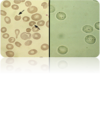
Q

The resulting mature RBC circulates for __ days, after which it is removed from the circulation by macrophages that detect senescent signals, primarily on the RBC membrane, through mechanisms that are poorly understood.

A

110 to 120 days

13
Q

Approximately ___ red blood cells are produced per hour in the bone marrow to maintain the hemoglobin level within fairly narrow limits.

A

10^10 (ten billion)

14
Q

Symptoms from anemia can result from two factors:

A

(1) decreased oxygen delivery to tissues, and, in patients with acute and marked bleeding, the added insult of (2) hypovolemia.

15
Q

Describe what happens with total blood volume in anemia

A

total blood volume remains normal in anemia due to chronic, low-grade bleeding, since there is plenty of time for equilibration within the extravascular space and renal retention of salt and water.

16
Q

How do tissues compensate for decrease oxygen delivery to tissues in anemia

A
  1. Increased SV 2. HR (therefore cardiac output) and 3. increased extraction of oxygen by the tissues .
17
Q

Primary symptoms of anemia

A
  1. exertional dyspnea 2. dyspnea at rest 3. varying degrees of fatigue** 4. signs and sx of hyperdynamic state (bounding pulses, palpitations, postural hypotension, roaring in the ears) 5. more severe: lethargy and confusion **Tend to be vague and non-specific
18
Q

PE findings of anemia

A

*Physical exam findings are poorly sensitive and specific with wide inter-observer differences. 1. pallor (face, nailbeds, conjunctiva, palms) 2. jaundice 3. Petechiae or bruising 4. LAN/HSM/sternal tenderness= concern for bone CA 5. tachy, dyspnea, fever, hypotension, *Stool should always be assessed for occult blood

19
Q

According to the kinetic approach, the following three questions are asked in order to determine the mechanism(s) causing the anemia.

A
  1. Is there evidence for decreased red cell production? 2. Is there evidence for increased red cell destruction (hemolysis)? –> jaundice 3. Is there a history of bleeding? (bright red blood in stool, mensturation, dark stools)
20
Q

Iron Deficiency Anemia (IDA) in the US is almost always secondary to ___

A

blood loss.

21
Q

types of hemorrhagic anemia (blood loss)

A
  1. Obvious Bleeding (e.g. trauma, melena, hematemesis, or menometorrhagia) 2. Occult Bleeding (e.g. Slow bleeding ulcer or carcinoma) Induced bleeding (e.g. 3. Hemodialysis, excessive donation, iatrogenic post surgical)
22
Q

What happens in hemolytic anemia

A

In hemolytic Anemia (Increased RBC Destruction), bone marrow is unable to keep up with the need to replace more than about 5% RBC Mass per day.

23
Q

Factors of hemolytic anemia

A
  1. Erythrocyte factors (memrane, surface protein, enzyme deficit, hemoglobinopathies) -inherited (sickle cell, thalassemia, spherocytosis) 2. Plasma Factors: -Ab, drug induced -autoimmune hemolytic anemia, thrombotic thrombocytopenic purpura, HUS 3. Mechanical heart valve 4. burns
24
Q

What are causes of decreased RBC production

A
  1. Nutritional: Lack of nutrients, such as iron, B12, folate. (dietary lack, malabsorption, or blood loss) 2. Marrow infiltration 3. bone marrow suppression 4. Low levels of Trophic Hormones which stimulate RBC Production: e.g. EPO (chronic renal failure), Thyroid (hypothyroidism), and androgens 5. Anemia of Chronic Disease / Infection – infectious, inflammatory, malignant disorders
25
Q

What is pernicious anemia

A

Decreased RBC production bc the intestines cannot absorb B12 appropriately

26
Q

What are drugs that cause macrocytic anemia

A
  1. Ethanol 2. Hydroxyurea – sickle cell 3. Methotrexate – RA 4. Trimethoprim – Abx 5. Zidovidine (HIV) 6. Chemotherapy
27
Q

What is considered macrocytic anemia

A

MCV >100fL

28
Q

What are nonmegaloblastic macrocytic causes of anemia

A
  1. Liver Disease (Common) 2. Myelodysplasia (Common) 3. Brisk reticulocytosis 4. Hypothyroidism 5. Multiple myeloma 6. Aplastic anemia
29
Q

What do you do when you realize MCV >100fL

A
  1. r/o drugs that cause macrocytic anemia 2. r/o nutritional causes (folate or B12) 3. consider nonmegaloblastic macrocytic causes
30
Q

Describe MCV of B12 and/or folate deficiency

A

HIGH MCV >105-110 *>115

31
Q

histological changes: 1. Peripheral blood smear: hypersegmented PMNs (>5 lobes) 2. macroovalocytes

A

macrocytic anemias

32
Q

What are associated findings w/ macrocytic anemia

A
  1. High serum LDH 2. mild indirect hyperbilirubinemia 3. MCV >100
33
Q

How can you differentiate between B12 and folate deficiency symptomwise?

A

*B12 and folate deficiency have identical symptoms with the exception of neuropsychiatric picture associated with B12 -Neurological symptoms seen only in severe B12 deficiency -Neuro symptoms are subacute combined degeneration of the nervous system: 1. Sensory peripheral neuropathy 2. Posterior and lateral column damage 3. Impaired mentation

34
Q

Causes of B12 deficiency

A

**MC= malabsorption 1. sprue or enteritis 2. Pernicious Anemia - lack of intrinsic factor due to gastric atrophy (COMMON) 3. Crohn’s Disease - disease of terminal ileum which is site of B12 absorption 4. Postgastrectomy- permanently at risk for B12 deficiency 5. Insufficient intake - B12 found only in animal products so watch for deficiency in vegetarians (normal storage 2 years) 6. Methionine synthase inhibition due to nitrous oxide anesthesia/abuse

35
Q

Causes of folate deficiency

A
  1. Insufficient intake - COMMON, diet lacking folate often coupled with alcohol induced block in folate metabolism 2. Chronic hemolysis 3. Pregnancy 4. Malabsorption - due to Dilantin or jejunal disease 5. Dihydrofolate reductase inhibition due to methotrexate Pyrimethamine
36
Q

How do you dx B12 or folate deficiency

A

Elevated serum homocysteine (both) and/or methylmalonic acid (B12 deficiency only) - high sensitivity and specificity 1. B12 - Check methylmalonic acid (MMA) level –>(a normal level make B12 deficiency extremely unlikely). 2. Folate - Check Homocysteine–> (Folate may be effected by dietary changes) 3. If B12 Deficiency - Consider serum anti-intrinsic factor antibodies if pernicious anemia suspected. -Schilling test is used to differentiate pernicious anemia from primary malabsorptive disorders.

37
Q

Causes of macrocytic anemia

A
  1. B12 def. 2. Folate def. 3. Liver dz 4. ETOH 5. hypothyroidism
38
Q

__ is used to differentiate pernicious anemia from primary malabsorptive disorders.

A

-Schilling test

39
Q

Describe the management of B12 and folate deficiency

A
  1. Appropriate replacement therapy 2. Expect reticulocytosis in several days 3. Lifelong monthly B12 injections for pernicious anemia 4. Folic acid supplements before/during pregnancy to prevent neural tube defects 5. Folic acid supplements for chronic hemolysis
40
Q

What is considered microcytic anemia

A

MCV <80fL

41
Q

What are common causes of microcytic anemia

A
  1. Fe deficiency 2. Lead 3. thalassemia 4. Early anemia of chronic dz
42
Q
  1. hypchromic 2. anisocytosis 3. poikilocytosis 4. pale small RBC w/ a scant rim of pink hgb 5. “pencil” shpaed cells
A

Fe deficiency microcytic anemia

43
Q

Describe the level of ferritin in anemia of chronic dz

A

typically normal or high

44
Q

how do you definitively dx iron deficiency anemia

A

absent Fe stores on Prussian Blue stain of marrow *You will see people order Serum Fe, Total iron binding capacity (TIBC), and % saturation

45
Q

Causes of iron deficiency anemia

A
  1. External Bleeding - GI, epistaxis, menorrhagia, etc 2. Hemoglobinuria from chronic intravascular hemolysis 3. Increased iron requirement - pregnancy, infants, preadolescents **You need to discover the cause –Caused by bleeding until proven otherwise!
46
Q

What is the management of iron deficiency anemia

A
  1. Ferrous sulfate 300mg TID on empty stomach (better absorbed) or with meals if GI intolerance (frequent) *Finite treatment trial (2-3 months) will help differentiate from ACD. 2. Reserve iron gluconate (IV) for those who cannot tolerate or absorb oral iron
47
Q

How to approach microcytic anemias

A
  1. R/O iron deficiency anemia–> (blood smear, serum Fe, transferritin % saturation, total iron binding capacity (TIBC)) 2. Evaluation with normal serum ferritin
48
Q

Causes of microcytic anemias with normal serum ferritin

A
  1. Thalassemic syndromes 2. Sideroblastic anemias
49
Q

Quantitative disorders of globin synthesis

A

Thalassemia

50
Q

Where are Thalassemias most common

A
  1. Mediterranean area, 2. the Middle East, 3. Southeast Asia, 4. Africa
51
Q

How do you dx Thalassemia

A

-microcytic anemia w/ normal serum ferritin *Hemoglobin electrophoresis is initial test of choice (is not 100% sensitive)

52
Q

Blood smear reveals: microcystosis out of proportion to degrees of anemia, Polychromasia*, target cells*, ovalocytes, and basophilic strippling

A

Thalassemia

53
Q

Clinical Forms of Thalassemia

A
  1. Minor (= Trait) - microcytosis and little to no anemia 2. Intermedia - more anemia, splenomegaly, occasional transfusion requirement 3. Major - severe anemia, frequent transfusions, and significant extramedullary hematopoiesis 4. Hydrops fetalis
54
Q

Management of Thalassemia

A
  1. Liberal use of transfusions with concurrent iron chelation therapy for multiply-transfused 2. Splenectomy for some with massive splenomegaly 3. Folic acid supplements 4. Allogenic marrow transplant for severely affected 5. Genetic counseling
55
Q

Most commonly includes defective heme synthesis in RBCs with abundant iron

A

Sideroblastic anemia

56
Q

In Sideroblastic anemia, Coarse granules of ferritin accumulate in mitochondria that encircle nucleus leads to ___

A

“Ringed Sideroblasts”

57
Q

Prussian blue stain of the bone marrow in a patient with refractory anemia. Blue-stained ferritin iron deposits in the mitochondria of erythroid precursors form an apparent ring around the nucleus (see arrows).

A

Sideroblastic anemia *also has ring sideroblasts (RARS)

58
Q

Causes of Sideroblastic anemia

A
  1. Genetic X-linked (low MCV) 2. Alcohol 3. Isoniazid and other anti-TB agents (low MCV) 4. Copper deficiency possibly due to zinc overload 5. Lead toxicity** 6. Myelodysplastic syndrome - refractory anemia with ringed sideroblasts, variable MCVs
59
Q

Peripheral blood smear shows basophilic stippling in several red cells from a patient with lead poisoning.

A

Sideroblastic anemia secondary to lead poisoning

60
Q

Management of sideroblastic anemia

A
  1. Removal of causative agents 2. Consider trial of pyridoxine therapy if genetic defect suspected
61
Q

What is considered normocytic anemia

A

MCV 80-100 **Remember early microcytic or macrocytic processes may present as normocytic anemia (MCV 80-100) OR simultaneous microcytic and macrocytic processes (iron deficiency and folate def in alcoholic) may present as normocytic anemia

62
Q

Causes/categories of normocytic anemia

A
  1. secondary anemia (anemia of chronic dz or hemolytic anemias) 2. Primary bone marrow failure
63
Q

Causes of secondary anemias leading to normocytic anemia

A
  1. Anemia of chronic disease: -chronic infections -Anemia of renal insufficiency (decreased EPO) -Anemia associated with chronic liver disease -Anemia associated with endocrinopathies: -Hypothyroidism -Addison’s Disease -Hypogonadism 2. Hemolytic Anemia’s -Sickle Cell Anemia -G6PD
64
Q

Causes of primary bone marrow failure leading to normocytic anemia

A
  1. Aplastic Anemia – stem cell injury associated with infection, toxins, medications 2. Myelodysplastic Syndromes e.g. Leukemia, lymphoma, myelofibrosis, Granulomous disease
65
Q

What should you consider ordering if you suspect primary bone marrow failure leading to normocytic anemia

A

Consider Peripheral Blood Smear +/- Marrow Biopsy

66
Q

What type of anemia is anemia of chronic disease (ACD)

A

Normo or microcytic

67
Q

ACD is caused by

A
  1. Inflammation (IBD, RA, DM, connective tissue disease, Chronic Infections, Malignancy) 2. Chronic Organ Failure 3. Elderly, 20% of individuals over 85 (Diagnosis of exclusion)
68
Q

What chronic organ failures can cause ACD

A
  1. Anemia of renal insufficiency (Cr >1.5mg/dL) - decreased EPO 2. Anemia associated with chronic liver disease 3. Anemia associated with endocrinopathies: -Hypothyroidism -Addison’s Disease -Hypogonadism
69
Q

What is the pathophysiology of ACD

A

Current understanding suggests a cytokine-mediated process that inhibits RBC production and interferes with erythropoietin production or function.

70
Q

Diagnosing IDA in presence of chronic disease can be difficult:

A
  1. Both IDA and ACD - low serum Fe and decreased transferritin % saturation 1. In ACD, Ferritin should be normal to Increased. (Ferritin is acute phase reactant, therefore best non-invasive to differentiate.) 3. Marrow Fe stores 100% in ACD since reflects defective iron reutilization 4. Expect ACD = MCV>75 4. Expect IDA = MCV reduced in proportion to degree of anemia (<75)
71
Q

What is the treatment of ACD

A
  1. Address the issue causing the anemia of chronic disease. 2. In severe anemia or effecting ADL’s, may consider transfusions or parenteral EPO (controversial if malignancy).
72
Q

Hemolytic anemia can either be __ or __

A

congenital or acquired (ie. antibody, drug)

73
Q

Congenital diagnostic approach to hemolytic anemia

A
  1. Enzymopathies – G6PD 2. Hemoglobinopathies – Sickle Cell 3. Membrane defects - spherocytosis, elliptocytosis
74
Q

Acquired diagnostic approach to hemolytic anemias

A
  1. Immune hemolysis (Cold agglutinin, Warm Autoantibodies, alloimmune)
75
Q

Nonimmune hemolytic anemia causes

A
  1. TTP, 2. HUS, 3. prosthetic heart valve, 4. vascular malformations, 5. DIC, 6. graft rejection, 7. vasculitis, 8. disseminated malignancy, 9. preeclampsia/eclampsia
76
Q

peripheral smear with Heinz body hemolytic anemia. Left panel: red cells with characteristic bite-like deformity (helmet cells*) and red cell fragments (ie. schistocytes)*. Right panel: Heinz body preparation which reveals the denatured hemoglobin precipitates.

A

Hemolytic anemia

77
Q

Associated lab findings with hemolytic anemia

A

Positive features: 1. Indirect hyperbilirubinemia 2. Reticulocytosis 3. Elevated serum LDH Decreased: 1. haptoglobin, 2. hemoglobinuria, and 3. hemosiderinuria (suggest intravascular rather than extravascular)

78
Q

Describe sickle cell anemia

A
  1. HbS mutation in single amino acid replacement in beta chain with substitution of normal glutamic acid with valine 2. Low oxygen or dehydration precipitates sickling and leads to complications called crises
79
Q

How do you diagnosis sickle cell anemia

A
  1. characteristic RBC morphology: sickle cells in HgbS and target cells in HgbSC
80
Q

What is sickle cell trait

A

(heterozygote) - benign condition detected by commonly used screening tests (sodium metabisulfite test) -Some pts develop symptoms with extreme hypoxic conditions -Other pts develop low grade hematuria due to high osmolality in the renal medulla promoting HgbS polymerization with secondary renal tubular cell injury

81
Q

Crises of Sickle Cell anemia

A
  1. Vasocclusive crisis 2. splenic sequestration 3. Aplastic crisis
82
Q

What are vasocclusive crises

A

1) Painful bone pain like aseptic necrosis of femoral head 2) Chest syndrome - fever, pulm infiltrates, hypoxia 3) renal damage 4) priapism

83
Q

aplastic crises can be due to

A
  1. transient infection, 2. medication, 3. toxin, 4. folate deficiency associated with marrow hypoplasia
84
Q

ACUTE management of Sickle cell anemia

A
  1. Hydration, oxygen, analgesia 2. Possible transfusion 3. Attention to infection
85
Q

LONG TERM management of Sickle cell anemia

A
  1. Folic acid supplementation 2. Hydroxyurea (Increases fetal Hgb) 3. Genetic counselling
86
Q

Who is usually affected by G6PD deficiency

A

*Common 1. variant in about 11% of African American and Mediterranean, 2. variant in about 3% of affected Caucasian populations *X-linked recessive Most Common African American Males

87
Q

G6PD Deficiency leads to

A

-decreased NADPH -Enzymopathies Causing Hemolytic Anemia

88
Q

Decreased NADPH in RBCs causes

A

poor defense against oxidizing agents leading to hemolytic anemia

89
Q

TX of G6PD deficiency causing hemolytic anemia

A

Avoid oxidant drugs Oxidizing agents: sulfa drugs, nirtofurantoin, primaquin, anti-TB drugs, fava beans, infection

90
Q

If MCV is <78 suspect

A
  1. Iron deficiency (blood loss or dietary) 2. lead intox. 3. chronic disease 4. Thalassemias 5. Hemoglobinpahties 6. sideroblastic anemia
91
Q

The initial approach to the diagnosis of anemia is to

A
  1. perform a complete history and physical examination 2. Complete blood count (CBC) with white blood cell differential, platelet count, reticulocyte count, and 3. an examination of the peripheral blood smear
92
Q

Most common causes of microcytic anemias

A
  1. Iron deficiency 2. thalassemias 3. anemia of chronic disease
93
Q

Most common causes of macrocytic anemias

A
  1. alcoholism 2. liver disease 3. folic acid and vit. B12 deficiency 4. myelodyplasia
94
Q

If you suspect Hemolysis from the patient’s history, physical examination, or examination of the peripheral blood smear (eg, sudden onset of anemia, jaundice, splenomegaly, presence of spherocytes or schistocytes or other red cell shape changes). It is confirmed by the finding of:

A
  1. increased levels of indirect bilirubin and 2. lactate dehydrogenase, and 3. low levels of haptoglobin.
95
Q

What from a pts history and physical exam can suggest hemolytic anemia

A
  1. sudden onset of anemia, 2. jaundice, 3. splenomegaly, 4. presence of spherocytes or schistocytes or other red cell shape changes
96
Q

The presence of abnormal cells in the circulation (eg, nucleated RBCs, blasts, atypical mononuclear cells) and/or abnormal increases or decreases in absolute counts for granulocytes, lymphocytes, monocytes, or platelets suggests that the anemia is part of a more complex hematologic disorder (eg, leukemia, aplastic anemia, myelodysplastic syndrome, myeloproliferative disorder). What should you do?

A

Consultation with a hematologist would be appropriate at this point

97
Q

-MCV <80 -low Fe -High TIBC -Low ferritin

A

iron deficiency–> determine cause

98
Q

-MCV <80 -low Fe -normal or low TIBC -normal or high ferritin

A
  1. ACD: infection, inflammation, or malignancy
99
Q

-MCV <80 -Normal to high Fe -Any TIBC -Normal to high ferritin

A
  1. Sideroblastic anemia (Siderocytes on peripheral smear) 2. Thalassemia (teardrop red cells or target cells)
100
Q

-MCV >100 -elevated homocysteine -elevated methylmalonate

A

B12 deficiency

101
Q

-MCV >100 -elevated homocysteine -normal methylmalonate

A

Folate deficiency