Anatomy & Physiology Flashcards
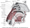
Whats considered Level 1 of the vocal tract?
is the vocal cord level. This is where ‘creak’, ’creaking’ ‘air added to the voice’, and ‘hammer vibrato’ are produced.
Whats considered Level 2 of the vocal tract?
is the level of the ventricular (false) folds. This is where ‘distortion’ is produced.
Whats considered Level 3 of the vocal tract?
is the level of the arytenoid cartilages/cuneiform, epiglottis and aryepiglottic folds. This is where ‘arytenoid rattle’, ‘saliva rattle’ and ‘growl’ are produced.
Whats considered Level 4 of the vocal tract?
is the level of the piriform fossa and posterior pharyngeal wall of the hypopharynx.
Whats considered Level 5 of the vocal tract?
is the level of the soft palate, uvula, back wall of the throat (oropharynx) and the back of the tongue. This is where the ‘uvula rattle’ and ‘back tongue rattle’ are produced.
Whats considered Level 6 of the vocal tract?
is the rest of the vocal tract (oral and nasal cavity). This is where the vowels and sound colour are produced. ‘Grunt’, ‘Scream’, ‘Vocal breaks’ and ‘Laryngeal vibrato’ are produced in a combination of various levels.
We gave the various parts in the vocal tract levels in order to identify and specify on which levels the various changes take place. The levels also make it easier to communicate where the changes take places.

Describe vocal cords
Some describe the vocal cord as consisting of two parts: 1) the membranous part anteriorly which is the part that vibrates and produces sound; 2) the cartilaginous or respiratory part posteriorly which is simply the mmucous membrane over the medial surface of the arytenoid cartilage.
Describe false cords
Also known as false cord or ventricular band. They are placed just above each vocal fold separated by a little crevice known as the ventricle. The false folds consist of outer lining of mucous membrane which encloses a band of fibrous tissue known as the quadrangular membrane. The false folds also contain fat and some muscle, the upper fibres throarytenoid muscle that insert into the epiglottis rather than the arytenoid cartilage sometimes known as the thyroepiglotticus.
Describe Arytenoid cartilages
These are paired pyramidal shaped cartilages that are attached to the sloping superior border of the posterior lamina of the cricoid cartilage via the cricoarytenoid joint by a complex of muscles and ligaments. The positioning of the arytenoid cartilages play an important role in regulating pitch as well as the opening and closing of the vocal folds. Each cartilage has an apex, a base, an anterolateral surface, a vocal process and lateral muscular process. The corniculate cartilage is attached to the apex. The base forms the upper surface of the cricoarytenoid joint. The vocal ligament is attached to the anterior vocal process while the quadrangular membrane is attached to the roughened anterolateral aspect. The posterior cricoarytenoid muscle, thyroarytenoid muscle and lateral cricoarytenoid muscles are attached to the muscular process. The interarytenoid muscle inserts into the posterior surface of the arytenoid cartilage which is triangular, smooth and concave.

Describe Cuneiform cartilages?
A piece of cartilage shaped like an ice cream cone placed in the posterior part of the aryepiglottic fold. They appear yellowy-white or cream coloured on endoscopic examination of the larynx. They are often mistaken for the arytenoid cartilages (they sit just in front of them). They probably help provide stiffness and control of the shape of the epiglottic funnel.

Describe Epiglottis
Flap of fibrocartilage that covers the entrance to the larynx helping prevent food and drink entering the trachea. It is one of nine cartilages that make up the larynx. The free upper part is broad and rounded. The lower, narrow part is attached to the angle formed by the two laminæ of the thyroid cartilage, a short distance below the superior thyroid notch, by the thyroepiglottic ligament. The lower part of its anterior surface is connected to the upper border of the body of the hyoid bone by an elastic ligamentous band, the hyoepiglottic ligament. The bulging posterior part of the inferior part of the epiglottis is known as the petiole. the epiglottis is normally pointed upward when breathing, speaking or singing rises during swallowing with the elevation of the hyoid bone moving to a more horizontal position and pointing backwards.
Describe the aryepiglottic folds
The aryepiglottic fold is a fold of mucosa forming the lateral border of the laryngeal inlet. It stretches from the apex of the arytenoid cartilage to the epiglottis. The corniculate cartilage and cuneiform cartilages lie under the surface of the free edge posteriorly stiffen and helping shape the upper rim of the epiglottic funnelknown as the laryngeal inlet. The fold contains the thin sheets of muscles: the aryepiglotticus and thethyroepiglotticus. The aryepiglottic fold forms the lateral border of the larynx separating it from the piriform fossa of the hypopharynx.

Describe the the piriform sinus

Also known as the piriform sinus (and sometimes wrongly spelt “pyriform”).The two piriform fossae (sinuses) are pear-shaped recesses on either side of the laryngeal inlet with the wider part anteriorly. Each has a medial wall formed by the aryepiglottic fold and an outer wall by the inner part of the thyroid cartilage and thyrohyoid membrane. There function is to provide channels for food to pass down during swallowing and the space within the fossa acts as a resonating chamber during speech and singing.
Describe the posterior pharyngeal wall

This is part of the hyopharynx. It forms the back wall of the throat from the level of the hyoid bone to the postcricoid region. It’s muscular walls are formed by the middle constrictor muscles which are attached anteriorly to the hyoid bone and stylohoid ligament and the thyrohyoid inferior constrictors which are attached anteriorly to the thyroid cartilage and cricoid cartilages forming a sling around the laryngopharynx.
Describe the hyopharynx

The hyopharynx is the funnel-shaped part of the throat on either side of the larynx stretching between the hyoid bone and joining the upper oesophagus at the level of the cricoid cartilage. It consists of three parts (subsites): the two piriform fossae, the posterior pharyngeal wall and the postcricoid region. Air passes through the upper part into the larynx and food also passes through the area into the oesophagus. Sound waves produced by the larynx resonate in this area to produce a dark sound colour. It is more open in the Neutral and Curbing and completely closed off in Edge.
Describe the soft palate
(or Velum palatinum) The fleshy movable part of the back of the roof of the mouth that closes off the nasopharynx during swallowing and speech. The central back part that hangs down is known as the uvula.
Describe the oropharynx
The oropharynx is part of the pharynx behind the oral cavity. It extends from the level of the soft palate to the level of the hyoid bone.
Describe the nasopharynx
the region between the soft palate and the back of the nasal cavities

What 3 parts does the pharynx consist of?
- nasopharynx the region between the soft palate and the back of the nasal cavities
- oropharynx the region between the soft palate and the hyoid bone
- hypopharynx, the region between the hyoid bone and the vocal folds.
Name the three important functions that the throat performs
- swallowing
- forming an air passage between the nose, mouth and larynx
- shaping of the vocal tract for voice.
Explain the Swallowing function
During swallowing it is of course important that food does not enter the windpipe. Once food has been chewed in the mouth the bolus is pushed backwards by the tongue into the back of the throat (oropharynx). To prevent the food bolus going into the nose the soft palate rises and the superior constrictor muscle contracts sealing off the nasopharynx. Together with the middle and inferior constrictor muscles it also helps propel the food bolus towards the oesophagus. The vocal folds and false cords close tightly together and the whole larynx rises upwards. The epiglottis tilts backwards forming a lid over the larynx preventing food going into the windpipe and helping direct it into the oesophagus. At the same time the upper oesophageal sphincter relaxes allowing the bolus to move from the pharynx into the oesophagus where a series of contractions propel it downwards into the stomach. Once the food has been swallowed the upper oesophageal sphincter closes off the top of the gullet so that food cannot be regurgitated back into the mouth. If you speak and eat at the same time food can easily go down the wrong way!

Explain the Air Passage function
Air can be breathed in through the nose or mouth. When passing through the nose the air is filtered from particles such as dust and pollen and also humidified and warmed. It also allows breathing while chewing food. However when large amounts of air are required such as during exercise it is necessary to breath through the mouth. We change between nose and mouth breathing subconsciously at rest or when talking. Mouth breathing tends to dry out the vocal cords a little more but it is sometimes necessary to get enough air in rapidly before singing the next phrase.
Explain shaping the vocal tract function
The voice is produced by the interaction of the sound produced by vibration of the vocal folds with the acoustic resonance of the vocal tract. Shaping the vocal tract by moving the lips, positioning the tonguein the mouth and oropharynx , moving the larynx up and down alters the vowel sounds and allows consonant sounds to be made for speech and singing. Lowering the larynx generally produces a darker sound colour while raising it causes a lighter sound colour. There are named constrictor muscles (superior, middle and inferior constrictor muscles) but there are many other ‘constrictor muscles’ in the neck such as the extrinsic laryngeal muscles which also help in the constriction and laryngeal positioning because of there attachment to the jawbone, base of the skull, sternum and collar bone. The intrinsic laryngeal muscles are the muscles within the larynx which open and close the vocal cords and alter the shape of the vocal folds so that the pitch, loudness and vocal mode can be changed.


