Anatomy of the Thorax Flashcards

1
Q

Describe the right main bronchis

A
  • Shorter
  • Wider
  • 2.5cm in length
  • Passes directly to the root of the lung at T5
  • Before joining the hilum, gives off upper lobe bronchus (this is not the case with the left main bronchus)
  • It then passes below the pulmonary artery to enter the hilum
2
Q

Describe the left main bronchis

A
  • Longer
  • More oblique
  • 5cm in length (hence twice the length of the right main bronchus)
  • Passes BELOW the Arch of the Aorta
  • Passes IN FRONT of the Oesophagus and descending aorta
  • Does not give off left upper lobe bronchus prior to the hilum, unlike the rigth main bronchus
  • Reaches hilum at T6 (left at T5)
  • Pulmonary artery spirals over left main bronchus: first lying anteriorly, then above it superiorly
3
Q

Views during bronchoscopy

A

Can visualise:

  1. Trachea
  2. Main bronchi
  3. Lobar bronchi
  4. Commencement of 1st segmental divisions
4
Q

Widening of the Trachea on CXR

A

Suggests enlargement of tracheobronchial lymph nodes

In the context of malignancy is a poor prognostic marker as lymph node involvement

5
Q

Origin of bronchial arteries

A
  • Bronchial arteries are branches ofteh descending thoracic aorta
  • They are of great clinical importance as they perfuse the lung parenchyma, hence during a PE the lung parenchyma is perfused despite pulmonary vessels being occluded
  • Supply each lobe of the lung parenchyma
6
Q

Drainage of the lung parenchyma

A

Bronchial veins drian the lung parenchyma

Bronchial veins drian into the azygous vein

7
Q

Drainages of the lung alveolar spaces

A

Oxygenated blood drains from the lung via the pulmonary veins

Superior and inferior pulmonary veins on each side

i.e. there are 4 pulmonary veins

Drains oxygenatedblood into theleft atrium

8
Q

Lymphatic drainage of the lungs

A
  1. Lymphatics of the lung drain centrapedally from the pleura to the hilum
  2. Bronchopulmonary lymph noes in the hilum drain to the tracheobronchial lymph nodes at the carina (enalrhgement causes splaying of the carina)
  3. Tracheobronchial lymph nodes then drain into the paratracheal lymph nodes
  4. Paratracheal lymph nodes drain into the mediastinal lymph nodes
  5. These mediastinal lymph nodes drain directly into the brachiocephalic veins or directly into the thoracic duct / right lymphatic duct
9
Q

Nerve supply to the lung

A

Innervation of the lungs is via the pulmonary plexus at the hilum

  • converys sympathetic fribres T2 - T5 (T6)
  • conveys parasympathetic fibres from the vagus nerve
10
Q

Constituents of a bronchopulmonary plexus

A

Consist of:

  1. A segmental artery
  2. A segmental vein
  3. A segmental bronchus

Wedge-shaped

Apices situated at the hilum and base at lung surface

If resected carefully –> little bleeding or air leak from raw surface

11
Q

Lingular segment

A

Left upper lobe has lingular segment

(= right middle lobe)

12
Q

Right upper lobe bronchopulmonary segements

A

Right upper lobe bronchopulmonary segements

  1. Apical bronchis
  2. Posterior bronchus
  3. Anterior bronchus
13
Q

Right middle lobe bronchopulmonary segements

A

Right middle lobe bronchopulmonary segements

  1. Lateral bronchus
  2. Medial bronchus
14
Q

Right lower lobe bronchopulmonary segements

A

Right lower lobe bronchopulmonary segements

  1. Apical bronchus
  2. Medial basal (cardial) bronchus
  3. Anterior basal bronchus
  4. Lateral basal bronchus
  5. Posterior basal bronchus
15
Q

Right bronchopulmonary segments

A

Upper

APA

Apical

Posterior

Anterior

Middle

LM

Lateral

Medial

Lower

AMALP

Apical

Medial basal

Anterior basal

Lateral basal

Posterior basal

16
Q

Left upper lobe bronchopulmonary segments

A

Left upper lobe bronchopulmonary segments

  1. Apicoposterior bronchus
  2. Apicoposterior bronchus
  3. Anterior bronchus
17
Q

Left middle lobe bronchopulmonary segments

A

Left middle lobe bronchopulmonary segments

  1. Superior bronchus
  2. Inferior bronchus
18
Q

Left lower lobe bronchopulmonary segments

A

Left lower lobe bronchopulmonary segments

  1. Apical bronchus

7.

  1. Anterior basal bronchus
  2. Lateral basal bronchus
  3. Posterior basal bronchus
19
Q

Left bronchopulmonary segments

A

Upper lobe

AA

Apicoposterior bronchus

Anterior bronchus

Lingula / middle

SI

Superior bronchus

Inferior bronchus

Lower lobe

AALP

Apical bronchus

Anterior basal bronchus

Lateral basal bronchus

Posterior basal bronchus

20
Q

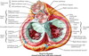
Defining the mediastinum

A

Cross-sectional midline of the mediastinum defined from the sternal angle anteriorly to the T4 vertebrae posteriorly

Above this is the superior mediastinum

Below this is the inferior mediastinum

The inferior mediastinum is divided into the anterior, middle and posterior divisions

Anterior: in front of fibrous pericardium

Middle: pericardium and great vessels

Posterior: from posterior surface of pericardium to T5 - T12 vertebral bodies

21
Q

Divisions of the inferior mediastinum

A

The inferior mediastinum is divided into the anterior, middle and posterior divisions

Anterior: in front of fibrous pericardium

Middle: pericardium and great vessels

Posterior: from posterior surface of pericardium to T5 - T12 vertebral bodies

22
Q

Fusions of the pericardium

A

Conical fibrous sace containing the heart and roots of the great vessels

Apex is fused with the adventitia of the great vessels

Base is fused with the central tendon of the diaphragm

23
Q

Anterior relations of the pericardium

(4)

A

Body of the sternum

Attached by the sternocardial ligaments

3rd - 6th costal cartilages

Anterior borders of the lungs

24
Q

Posterior relations of the pericardium

(6)

A
  1. Oesophagus
  2. Descending aorta
  3. T5 - T8 verebrae
  4. Roots of the lungs
  5. Mediastinal pleural
  6. Phrenic nerve
25
Q

Define the pericardial cavity

A

The potential space between the parietal pleura and visceral pleura of the pericardium

26
Q

Define the pericardial reflections

A

The pericardial pleura is reflected around the roots of the great vessels

-the parietal pleura becomes continuous with the the viscera pleura / epicardium

Marked on the posterior surface by:

OBLIQUE sinus: bound by IVC and four pulmonary veins and forms recess between left atrium and pericardium

TRANSVERSE sinus: SVC and left atrium behind and the aorta and the pulmonary trunk in front (forms gap between veins and arteries)

27
Q

Define the oblique sinus

A

OBLIQUE sinus: bound by IVC and four pulmonary veins and forms recess between left atrium and pericardium

28
Q

Define the transverse sinus

A

TRANSVERSE sinus: SVC and left atrium behind and the aorta and the pulmonary trunk in front (forms gap between veins and arteries)

29
Q

Position of the heart

A

The herat is situated in the middle of the inferior mediastinum

The inferior mediastinum is defined by the T5 - T12 vetrebral bodies

The middle inferior mediastinum begns at the fribous pericardium

30
Q

Borders of the heart

A

Rigth border = right atrium

Left border = left venticle and left auricular appendage

Inferior border = Mianly right ventricle and lower rigth atrium with the apex of the left ventricle

Apex = left ventricle

31
Q

Surfaces of the heart

A

Anterior surface: mainly right ventricle, separated from right atrium by atriovententricular groove, separated from the left ventricle by the anterior interventricular grrove

Inferior (diaphragmatic) surface: left ventricle and rigth ventricle separatesd by the posterior interventricular groove

Base / Posterior surface: mainly left atrium and some of right atrium (quadrilateral in shape)

32
Q

Vessels draining into the rigth atrium

(4)

A
  1. SVC in the upper and posterior part
  2. IVC in the inferuir part
  3. Coronary sinus in the lower part
  4. Anterior cardiac vein in the anterior part (drains much of the anterior surface of the heart)
33
Q

Define the crista terminalis

A

Musuclar ridge running almost vertically down from the SVC to the IVC

Indicated on the outer surface of the heart by a shallow groove - the sulcus temrinalis

Separates the smooth walled posterior atrium derived from the sinus venosus from the rough-walled anterior atrium which is derived from the fetal atrium

34
Q

Define the sulcus terminalis

A

The sulcus terminalis is a shallow groove on the exterior surface of the heart that marks the inner crista terminalis

crista terminalis- a muscular ridge running from the SVC to the IVC

in the right atrium

35
Q

Define the pectinate muscles

A
  • musculi pectinati*
  • =* pectinate muscles

Trabeculations in the rougth anterior portion of the atrium (derived from fetal atrium) which are prodiuced by parallel columns of muscles

36
Q

Inflow and outflow of right ventricle

A

Joined by the right atrium via the vertically orientated tricuspid valve

Houses the pulmonary valve which leads to the pulmonary trunk

Infidibuloventricular crest is a muscular ridge that separates the inflow and outflow tracts of the right ventricle

37
Q

Define the infidibuloventricular crest

A

Infidibuloventricular crest is a muscular ridge that separates the inflow and outflow tracts of the right ventricle

Lies between the atrioventricular and pulmonary orifices

38
Q

Define the infundibulum (also known as conus arteriosus)

A

Infundibulum (also known as conus arteriosus) is a conical pouch formed from the upper and left angle of the right ventricle in the chordate heart, from which the pulmonary trunk arises.

39
Q

Define the trabeculae carnae

A

Irregular muscular elevations that mark the inflow path of the rigth ventricle

-from which, the papillary muscles project into the lumen and attach to the free borders ofthe tricuspid valve via the chordae tendinae

40
Q

Define the chordae tendinae

A

Chordae tendinae: tendon-resembling fibrous cords of connective tissue that connect the papillary muscles to the tricuspid valve and the mitral valve in the heart

They arise from the trabeculae carnae

41
Q

Action of the papillary muscles

A

During systole the papillary muscles shorten

–> pull upon the chordae tendinae

–>prevent prolapse of the tricuspid valve into the atrium

Ischaemic injury to the papillary muscles can precipitate acute valve failure and subsequent acute heart failure

42
Q

Define the moderator band

A

Moderator band: a muscular band that crosses the ventricular cavity from the interventricular spetum to the anterior wall

Contains: RIGHT branch of the ATRIOVENTRICULAR BUNDLE and carries it to the RIGHT VENTRICLE

43
Q

Infundibulum (of the heart)

A

= outflow tract of the rigth ventricle

Smmoth-walled, directed upwards and to the tight toward sthe pulmonary trunk

44
Q

Describing the left atrium

A
  1. Smaller vs the right atrium
  2. Thicker walled
  3. Four pulmonary veins open into the posterior aspect
  4. Fossa ovalis: shallow depression on the septal surface of the left atrium
  5. Main part of the atrium is msooth walled
  6. Auricular appendage contains ridges from the undelrying pectinate muscles
45
Q

Describing the left ventricle

A
  1. Communicates with the left atrium via the mitral valve: bishops mitre - attached to papillary muscle via the chordae tendinae
  2. Marked by thick trabeculae carnae
  3. The only smooth part of the left ventricle is teh fibrous vestibule below the aoritc orifice
46
Q

Describing the mitral valve

A
  • Separates the left atrium and teh left ventricle
  • Two leaflets: bishops mitre
  • Large anterior leaflet
  • Smaller posterior leaflet
  • Attahced by chordae tendinae by the papillary muscles
47
Q

Describing the aortic orifice

A
  • Circular opening, in front and to the right of the left atrioventricular orifice
  • Situated above aortic vestibule which is smooth-smalled
  • Three semi-lunar cusps = aortic valve
  • Above which there are three sinuses - the aortic sinuses
48
Q

Defining the aortic sinuses

A

Aortic sinus = dilitations in the ascending aorta

  1. Anterior aortic sinus: gives off rigth coronary artery
  2. Left posterior aortic sinus: gives off the left coronary artery
  3. Right posterior aortic sinus: non-coronary sinus

During diastole turbulent blood forms inthe sinuses which helps to shut the aortic valve and also perfuses the coronary arteries

49
Q

Location of the sinoatrial node

A

Located in the upper part of the crista terminalis (muscular ridge from SVC to IVC in rigth atrium)

50
Q

Location of the atrioventricular node

A
  • Located in the atrial septum immediately above the opening of the coronary sinus
  • Bundle of His divides at the junction ofthe membranous and muscular parts of the interventricular septum
51
Q

Describing teh conduction system of the heart

A
  1. Sinoatrial node is located in the upper part of teh crosta terminalis, just to the right of the opening of the SVC and initiates the electrical impulse
  2. From here, the impulse spreads through the atrial musculature to the atrioventricular node
  3. The atrioventricular node is lcoated in the atrial septum just above the coronary sinus
  4. It continues as teh Bundle of His which divides at the junction of the membranous and muscular parts of the interventricular septum
  5. The rigth and left branches runs immediately beneath the endocardium
52
Q

Origin of the rigth coronary artery

A

= ANTERIOR AORTIC SINUS

53
Q

Origin of the left coronary arery

A

= LEFT POSTERIOR AORTIC SINUS

54
Q

Path of the right coronary artery

A
  1. Arises from the anterior aortic sinus
  2. Passes forwards (anteriorly) between the pulonary trunk and the right atrium
  3. Descends in the right section of the atrioventricular groove
  4. At the inferior border of the heart it continues along the atrioventricular groove to anastomose with the LEFT CORONARY ARTERY at the posterior interventricular groove

Branches

  • Gives off marginal artery along the lower border of the heart
  • Gives off posterior interventricular branch at the posterior aspect which runs forward in the interventricular groove to anastomose near the apex with corrosponding branch of LCA
55
Q

Path of the left coronary artery

A
  1. Arises from the left posterior aortic sinus
  2. Passes behind and then to the left of the pulmonary trunk
  3. Reaches left part of the atrioventricular groove in which it runs laterally around the heart as the cirucmflex artery
  4. Circumflex artery reaches the posterior interventricular groove

Branches

  • Anterior interventricular artery aka Left Anterior Descending: given off within 2.5cm of its origin, supplies the anterior aspect of both the left and right ventricles, passes around the apex of the heart to anastomose with the posterior interventricular (or posterior descending) branch of the RCA
  • This bifurcation into LAD and circumflex can occassionalyl be a trifurcation with an additional artery called the ramus or intermediate artery being given off
  • The circumflex gives off the left marginal artery
56
Q

Branches of the rigth coronary artery

A

Branches

Gives off marginal artery along the lower border of the heart and continues as the rigth coronary artery

In 85% of people it then gives off the posterior descnding artery (posterior interventricular branch) at the posterior aspect which runs forward in the interventricular groove to anastomose near the apex with corrosponding branch of LCA

57
Q

Branches of the left coronary artery

A

Left main stem --> left anterior descending ( anterior interventricular artery) and left circumflex

Left anterior descending (anterior atrioventricular artery) –> diaganol artery

Left circumflex –> left merginal artery

Anterior interventricular artery aka Left Anterior Descending: given off within 2.5cm of its origin, supplies the anterior aspect of both the left and right ventricles, passes around the apex of the heart to anastomose with the posterior interventricular (or posterior descending) branch of the RCA

This bifurcation into LAD and circumflex can occassionalyl be a trifurcation with an additional artery called the ramus or intermediate artery being given off

The circumflex gives off the left marginal artery

58
Q

Venous drainage of the heart

(3)

A
  1. Coronary sinus
    Great cardiac vein
    Middle cardiac vein
    Small cardiac vein
    Oblique vein
  2. Anterior cardiac veins
  3. Venae cordis minimi
59
Q

Describe the coronary sinus

(4)

A
  • The coronary sinus lies in the posterior atrioventricular groove
  • It opens up into the rigth atrium just to the left of the IVC
  • Recieves
  1. Great Cardiac Vein (fed by the anterior interventricular vein): in the anterior atrioventricular groove
  2. Middle Cardiac Vein: in the inferior atrioventricular groove
  3. Small Cardiac Vein fed by the rigth marginal vein: in the lower rigth border of the heart accompanying the right marginal artery
  4. Oblique Vein: descends obliquely in the posterior aspect of the left atrium
60
Q

Describe the anterior cardiac veins

A

Cross the anterior atrioventricular groove and open up diretcly into the anterior surface of the rigth atrium

Up to three or four in number

61
Q

Describe the venae cordis minimi

A

Small veins that drain directly into teh cardiac cavity

62
Q

Nerve supply to the heart

A

Vagus nerve: parasympathetic supply that reduces heart rate

Cervical and T1 - T5 vertebrae: sympathetic supply via the superficial and deep cardiac plexus which increases heart rate

Referred pain: to neck / arm via the T1 - T5 afferent sympathetic nerves

63
Q

Components of the primitive heart tube

(5)

A
  1. ​Truncus arteriosus (cephalic)
  2. Bulbus cordis
  3. Ventricle
  4. Atrium
  5. Sinus venosus (caudal)
64
Q

Describe the primitive heart

A
  • 5 dilatations along primitive heart tube forms into adult heart structures
  • Dextral looping of the primitive heart aligns the heart chambers and structures
  • Septum and valves develop which allow for separation of the venous and arterial circulatory pathways

5 dilitations

cephalic

  1. Truncus arteriosus
  2. Bulbus cordis
  3. Ventricle
  4. Atrium
  5. Sinus venosus

caudal

65
Q

Brief overview of the embryological development of the heart from the heart tube

A
  1. The primitive heart tube elongates
  2. Kinks
  3. Caudal end recieiving blood from the sinus venosus comes to lie behind the cephalic end
  4. Sinus venosus absorbs into the atrium
  5. Bulbus cordis absorbs into the ventricles
  6. Result of which is the atria and great veins lie behind the ventricles
66
Q

Division of the primitive atrium

A

Septum primum: grows downwards from the posterior and superior walls of the primitive common atrium - hole appears in upper part prior to fusion at lower border

Septum secundum: second membrane to the right of the septum primum BUT remains incomplete - free lower edge

67
Q

Septum primum

A
  • Grows downwards from the posterior and superior walls of the primitive common atrium
  • Fuses with the endocardial cushions
  • Prior to complete fusion, a hole appears in the upper part of the septum = foramen secundum (in the septum primum)
68
Q

Foramen secundum

A

= hole in the upper part of the septum primum

69
Q

Septum secundum

A
  • Second memrbane to the right of the septum primum
  • Grows downwards
  • Incomplete growth and has free lower border
  • Hence does not fuse with endocardial cushions
  • Overlaps foramen secundum forming a valve-like structure that allowes blood to enter from the rigth atrium to the left atrium = FORMAN OVALE
70
Q

Hole in the upper part of the septum primum

A

Foramen secundum

71
Q

Valve-like structure composed of the septum primum and septum secindum overlapping the foramen secundum

A

=Foramen ovale

Allows bloods from the rigth atrium to enter the left atrium

72
Q

Proportion of adults with patent foramen ovale

A

10% have anatomically patent (but functionally closed) foramen ovale

73
Q

Division of the primitive ventricles

A
  1. Up-growth of septum from apex of the heart
  2. Stops short if the endocardial cushiosn creating interventricular foramen
  3. Single truncus arteriosus divides into the aorta and pulmonary trunk by the SPIRAL SEPTUM
  4. Spiral septum continues growing down into the ventricles
  5. Completes the division of the ventricles by forming the pars membranacea septi = small upper part of interventricular septum
74
Q

Fusions of the developing atria

A

1. Primitive sinus venosus fuses with Atrium: smooth walled part of the atrium in developed heart is the contributon from the sinus venosus , pectinate part is from the primitive atrium and forms the auricular appendage

2. Pulmonary venous trunk is absorbed into the left atrium: the pulmonary venous trunk forms the smooth-walled part of the developed left atrium, trabeculated part if from the primitive atria and forms the auricular appendage

75
Q

pars membranacea septi

A

= continuation of the sprial septum forming the upper part of the interventicular septum, the spiral septum progresses down it divides the single truncus arteriosus into the aorta and the pulmonary trunk

Completes interventricular spetum to form left and rigth ventricles

76
Q

Number of aoritc arches

A

= SIX PAIRS

77
Q

Site of origin of aortic arches

A

Six aortic arches (pairs) arise from the truncus arteriosus

78
Q

Aortic arches that DISSAPEAR

A

1st pair

2nd pair

5th pair

3 pairs dissapear, 1st, 2nd and 5th

79
Q

Development of 4th aortic arches

A

4th Aortic Arches

RIGHT: Brachiocephalic and subclavian arteries

LEFT: Arch of the aorta

HENCE:

Distal sixth arch on the rigth dissapears, next arch is the 4th

Distal sixth arch perisst as the ductus arteriosus on left

Recurrent laryngeal nerve is caught on the 4th arch on the rigth which is the subclavian artery,

Caught on the sixth arch on the left with ends up being the ligamentum arterosum

80
Q

Development of the 3rd aortic arches

A

3rd Aortic Arches

RIGHT and LEFT: become carotid arteries

81
Q

Development of 6th aortic arches

A

6th Aortic Arches

RIGHT: proximal part forms the right pulmonary artery

LEFT: proximal part forms the left pulmonary artery and the distal part forms ductus arteriosis

82
Q

Development of the vagus nerve

A
  1. Vagus nerve lies lateral to the primitive pharynx and is separated from it by the aortic arches
  2. The vagus nerve passes medially caudal to the aortic araches to supply the developing larynx
  3. Elongation occurs, the developing heart and aoritc arches elongate caudually catching the vagus nerve
  4. Right DISTAL 6th aortic arch dissapears, right recurrent laryngeal next caught on the 4th aortic arch which is the rigth subclavian artery
  5. Left DISTAL 6th aortic arch becomes the ductus arteriosus and hence the left recurrent laryngeal nerve is wrapped around the ligamentum arteriosum
83
Q

Vertebral level of the superior angle of the scapula

A

T2

84
Q

Vertebral level of the suprasternal notch

A

T2/T3

85
Q

Vertebral level of the sternal angle

A

= Angle of Louis

= maunbrosternal junction

= second costal cartilage

= T4/ T5

86
Q

Vertebral level of the inferior border of the scapula

A

Inferior border of the scapula as it overlies the 7th ribs is at the level of T8

87
Q

Vertebral level of the xiphisternal joint

A

=T9

88
Q

Vertebral level of the subcostal line

A

Lower part of the costal margin at the 10th rib = subcostal line

Passes through L3

89
Q

Describing the manubrium

A

Manibrium is latin for handle

Overlies aortic arch and corrosponds to T3-T4 vertebra

Manubrium is joined ot the body of the sternuma at the angle of luis

90
Q

Angle of Louis

A

= second costal cartilage

= T4 / T5

91
Q

Bony prominences of the thorax

(6)

A

Superior angle of the scapula = T2

Upper border of the manubrium / suprasternal notch = T2/T3

Sternal angle = T4/T5

Inferior border of the scapula as it overlies 7th rib = T8

Xiphisternal joint = T9

Subcostal line (at costal margin of 10th rib) = L3

92
Q

First palpable spinous process

A

=C7

C7 is the only cevrical vertebrae to have a significant spinous process (the C1- C6 vertebrae have bifid spinous processes)

93
Q

Definition of apex beat

A

=Most inferior and lateral palpable heart beat

Normally: 5th intercostal space 9cm from the midline (midclavicular line)

94
Q

Location of male nipple line

A

4th intercostal space

95
Q

Levels of the trachea

A
  • Trachea commences at the cricoid cartilage at C6
  • Runs vertically downards and ends at the level of the sternal angle at T5 just to the rigth of the midline
  • In the erect position in full inspiration the trachea bifurcates at the level of T6
96
Q

Limits of the cervical pleura

A

Rises higher than expected and can be injured causing pneumothorax (a classic case of this is during insertion of subclavian central line and subsequent respiratory decline)

Defined by a curved line drawn from the sternoclavicular joint to the junction between the medial third and middle third of the clavicle

Rising 2.5cm above the clavicle in a dome-shape

97
Q

Levels of the pleural lung markings

A
  1. Highest point is the reaching from the sternoclavicular joint to the junction of the first thrid and middle third of the clavicle
  2. Passes to the midline at the Angle of Louis
  3. Descends to the 6th costal cartilage
  4. Then begins to pass medially
  5. Crosses the 8th rib in the mid-clavicular line
  6. Crosses 10th rib at mid-axillary line
  7. Crosses 12th rib at the lateral border of the erector spinae muscle
  8. At medial extremity it extends to just below the 12th rib margin

On the left side it runs away from midline to lateral sternal border at 4th costal cartilage

98
Q

Loin excision causing pneumothorax

A

You can accidentally open the pleura with a loin incision (daining subphrenic abscess or exposing kidney)

As the upper incison curves eitehr towards the 10th rib at the costal margin or if it extends posteriorly to infring upon the pleura as it crosses the 11th / 12th rib

99
Q

Surface markings of the lungs

A

Apex of both lungs follows the contours of the pleura

-sternoclavicular joint to junction of first thrid and middle third of the clavicle

Right lung follows contour of mediastinal pleura

Left lung has distinct cardiac notch and passes behind the 5th and 6th costal cartilages

Lower borders, crosses:

6th rib mid-clavicular line

8th rib mid-axillary line

10th rib adjacent to vertebral column

i.e. two rib spaces above the pleura

100
Q

Anatomy of the oblique fissure

A

Runs from 2.5cm lateral to the spine of T3

Along the 5th intercostal space

Runs to to the 6th costal cartilage 3cm from the midline

There is an oblique fissure on each side

101
Q

Anatomy of the horizontal fissure

A

Continues as a horizontal line along the 4th costal cartilage (anteriorly heading backwards)

Meets oblique fissure as it cross the 5th rib

Only present in the RIGHT lung

102
Q

Surface markings of the heart

A

RIGHT SIDE:

3rd costal cartilage 1/2 inch form the sternum

6th costal cartilage 1/2inch from the sternum

LEFT SIDE:

2nd COSTAL CARTILAGE 1/2 inch from sternum

5th intercostal space 3.5 inch from the MIDLINE

103
Q

Avoiding the internal thoracic artery

A
  • The internal throacic arteries (internal mammary arteries) run vertically behind the costal cartillages ~ 1.25cm lateral from the sternum
  • They should be avoided in aspiration procedures of the lung as they can bleed dramatically
104
Q

Number of ribs

A

12 pairs

7 pairs that articulate with the sternum directly

ribs 1 - 7

3 pairs that articular with the sternum via the costal cartilage above

ribs 8 - 10

2 pairs of floatig ribs

ribs 11 -12

105
Q

Attachment of the costotransverse ligament

A

Attaches to the stout neck of the rib

Lateral costotransverse liagment - at level of vertebrae

Superior costotransverse ligament - from vetrebrae above

106
Q

Head of the rib

A

Two facets

Superior facet: joins with the LOWER demifacet of the vertebrae BELOW

Inferior facet: joins with the UPPER demifacet of the vertebrae BELOW

107
Q

Tubercle of the rib

A

smooth facet for articulation with the transverse prcoess of the named vertebrae at that level

108
Q

Site of attachement of the lateral limit of erector spinae muscle

A

= angle of the rib

109
Q

Properties of the first rib

(6)

A
  • Flattended (from above –> downards)
  • Prominent tubercle on inner border for attachment of anterior scalene muscle
  • Groove for suubclavian artery
  • Groove for subclavian vein
  • Single articular facet
  • Shortest
110
Q

Clinical use of subclavian groove

A

Subclavian artery and lowest trunk of brachial plexus lie behing anterior scalene muscle

Site of anesthetic infiltration for the brachial plexus into the subclavian groove

Most of the surface of clavicle is occupied by a groove, which gives attachment to the Subclavius; the coracoclavicular fascia, which splits to enclose the muscle, is attached to the margins of the groove.

111
Q

List the atypical ribs

A

1st, 10th, 11th and 12th

Rib 1 is shorter and wider than the other ribs. It only has one facet on its head for articulation with its corresponding vertebrae (there isn’t a thoracic vertebrae above it). The superior surface is marked by two grooves, which make way for the subclavian vessels.

Rib 2 is thinner and longer than rib 1, and has two articular facets on the head as normal. It has a roughened area on its upper surface, from which the serratus anterior muscle originates.

Rib 10 only has one facet – for articulation with its numerically corresponding vertebrae.

Ribs 11 and 12 have no neck, and only contain one facet, which is for articulation with their corresponding vertebrae

RIb 11: shallow sibcostal groove, slight angle

Rib 12: no groove, no angle

112
Q

Weakest point of a rib

A

Criush injuries often cause fractures close to the angle of the rib as this the ribs weakest point during AP compression

113
Q

Notching of the ribs

A

Roesler’s sign = enlarged collateral vessel

Seen in:

  • co-arctation of the aorta
  • interrupted aortic arch
  • subclavian artery obstruction
  • AVM
  • Tetrallogy of fallot
114
Q

Collateral vessels in co-arctation of the aorta

A

Co-arctation means that the aorta beneath the narrowing would not recieve blood

The internal throacic artery (internal mammary) branches prior to the co-arctation and passes vertically behind the costal cartillages

Anterior intercostal branches of the internal throacic artery are connected to the intercostal arteries

These intercostal arteries are a direct branch of the descending aorta

Increased flow from the internal throacic –> anterior intercostal branches –> intercostals –> descending aorta

Increased blood flow down inferior epigastric artery too

Produces Roesler’s sign (Notching of the ribs)

115
Q

Cervical ribs

A
  • 0.5% of the population
  • 50% of cases are bilateral
  • Attachment ot the transverse process of C7
  • Articulate with the first rib
  • Some are truncated and have fibrous band connetcign to the first rib
  • Cause pressure on lowest trunk of the brachial plexus –> paraesthesia ober ulnar border of forearm and wasting of hand muscle (T1)
  • Can also cause post-stenotic dilatation of subclavian artery
116
Q

Fractures of the sternum

A
  • Elastic costal cartlages means that fractures of the sternum are rare
  • Associated with fracture-dislocations of the thoracic spine
117
Q

Blalock-Taussig Shint

A

Procedure: length of artificial tubing, 3 to 4 millimeters in diameter, is sewn between either the subclavian or the carotid artery and the corresponding side branch of the pulmonary artery

Indication: Increase blood flow in pulmonary artery in cyanotic heart defects such as tetrallogy of fallot or pulmonary atresia

118
Q

Direction of external intercostal muscles

A

External intercostals run forwards and downards

From rib above –> to rib below

Run from vertebare to costochondral junctionalm at the costsochondral junction teh muscle is replaced by the anterior intercostal memrbane

119
Q

Direction of internal intercostal muscles

A

Run backwards and downwards

Becomes the posterior intercotsal membrane

120
Q

Intercostal membranes

A

External intercostal aponeurosis = anterior intercostal membrane at point of costochrondral joint

Internal intercostal aponeurosis = posterior intercostal membrane

121
Q

Innermost intercostal muscles

A

Continuous with the internal intercostals

Spans multiple intercostals at a time

Anteriorly is more distinct as the transversus thoracis muscle

122
Q

Origins of the posterior intercostal arteries

A

1st and 2nd: branches of superior intercostal artery, which is a branch of the costocervical trunk, which is the only branch of the second part of the subclavian artery

3rd - 12th: direct branches of the descending aorta

Each posterior intercostal artery branches to skin, muscle AND spinal cord

123
Q

Origin of anterior intercostal arteries

A

1st - 6th: Direct branches of internal throacic artery

These anterior intercostal arteries give rise to the perforatingbranches for spaces 1 -6

7th - 9th: Branches of the musculophrenic artery which a is branch of the internal thoracic artery

The bottom two spaces only have posterior intercostal arteries

124
Q

Origin of the intercostal nerves

A

Anterio rami of the thoracic nerves

Each gives off :

  1. muscular branch
  2. lateral cutaneous branch
  3. anterior cutaneous branch
125
Q

Intercostal nerve block

A

Local anaesthetic is infiltrated around the intercostal nerve trunk

-arises from anterior primary rami of each thoracic nerve

Needle injected on posterior back around angle of rib

126
Q

Posterolateral thorocotomy

A

Incision along the line of the 5th or 6th rib

127
Q

Nerve supply to the chest wall

A

Lateral cutaneous branches (rami cutanei laterales) are derived from the intercostal nerves, about midway between the vertebræ and sternum; they pierce the Intercostales externi and Serratus anterior, and divide into anterior and posterior branches.

The anterior branches run forward to the side and the forepart of the chest and skin, fourth nerve anterior branches supplying the areola and the mamma; those of the fifth and sixth nerves supply the upper digitations of the Obliquus externus abdominis.

The posterior branches run backward, and supply the skin over the scapula and Latissimus dorsi.

128
Q

Distribution of intercostal nerves

A

Intercostal nerves 1 -2: supply fibers to the upper limb in addition to their thoracic branches

Intercostal nerves 3 - 6 : limited in their distribution to the walls of the thorax

Intercostal nerves 7- 11 : supply the walls of the thorax and abdomen.

7th intercostal nerve terminates at the xyphoid process, at the lower end of the sternum.

10th intercostal nerve terminates at the navel.

12th (subcostal) thoracic is distributed to the abdominal wall and groin.

129
Q

Description of the diaphragm

A

Diaphragm is a dome-shaped septum that separates the thoracic and abdominal cavities and plays a role in respiration.

It is made up of two portions: a peripheral muscular portion and a central tendinous aponeurosis

It has three main openings for the IVC, oesophagus and aorta

130
Q

3 constituent parts of the muscular portion of the diaphragm

A
  1. Vertebral part: right and left crura, medial arcuate ligament, lateral arcuate ligamet and median arcuate ligament
  2. Costal part: attached to inner aspect of lower six ribs and costal cartilages
  3. Sternal portion: small muscular slips from deep surfae of xiphisterum
131
Q

What is the medial arcuate ligament?

A

Arcuate ligament refers to the fibrous arches formed by the thickening of the fascia.

The medial arcuate ligament is a thickening of the fascia of the psoas major muscle.

132
Q

What is the lateral arcuate ligament?

A

Arcuate ligament refers to the fibrous arches formed by the thickening of the fascia.

The lateral arcuate ligament is a thickening of the fascia of the quadratus lumborum muscle, which makes sense as this is lateral to the psoas major which makes up the medial arcuate ligament.

133
Q

What is the MEDIAN arcuate ligament?

A

The median arcuate ligament is the fusion of the two medial borders of the left crus and right crus in front of the aorta

Right crus: L1 - L3

Left crus: L1 - L2

Can cause compression of ceoliac artery, causing MALS

134
Q

Innervation of the diaphragm

A

Phrenic nerve

C3, C4, C5

Long course of phrenic nerve follows embryological course of diaphrgam

135
Q

Referred pain by the phrenic nerve

A

The sensory nerve fibres from the central part of the diaphragm also run
in the phrenic nerve; hence, irritation of the diaphragmatic pleura (in pleurisy) or of the peritoneum on the undersurface of the diaphragm by subphrenic collections of pus or blood produces referred pain in the corresponding cutaneous area, the shoulder‐tip.

136
Q

Sensory supply to the diaphragm

A

Peripheral part: intercostals

Central tendon: phrenic nerve

137
Q

Openings in the diaphragm

A

IVC: T8

  • situated in central tendon, slightly right to midline
  • also site of transmission for right phrenic nerve

Oesophagus: T10

  • situated in muscular fibres of left and rigth crura slightly to left of midline
  • transmits left gastric artery and vein
  • trasnmits two vagus nerves

Aorta: T12

  • lies in midline
  • tansmits throacic duct and azygous vein

Left phrenic nerve pierces diaphragm alone

Greater and lesser splanchnic nerves pierce the crura

Sympathetic chain passes behind the diaphragm deep to the medial arcuate ligament to reach the posterior abdominal wall

138
Q

Level of IVC entering diaphragm

A

IVC: T8

  • situated in central tendon, slightly right to midline
  • also site of transmission for right phrenic nerve
139
Q

Level of aorta entering diaphragm

A

Aorta: T12

  • lies in midline
  • tansmits throacic duct and azygous vein
140
Q

Level of oesophagus entering diaphragm

A

Oesophagus: T10

  • situated in muscular fibres of left and rigth crura slightly to left of midline
  • transmits left gastric artery and vein
  • trasnmits two vagus nerves
141
Q

Diapgramatic apertures

A
142
Q

Embryological components of the diaphragm

A

COME BACK TO

143
Q

Symptoms of sliding hiatus hernia

A

Cardia of stomach is above the hiatus.

Hence reguritation of peptic juices is common.

Leads to:

  • heart burn
  • oesophagitis
  • bleeding
  • stricture formation
144
Q

Symptoms of rolling hiatus hernia

A

Rolling hiatus hernia are also known as para-oesophageal hernia.

The cardia remians beneath the diaphragm but the fundus rises above it.

There is little regurgitation as the cardia is below the hiatus, cardio‐oesophageal junction is intact.

Leads to:

  • epigastric discomfort
  • flatulence
  • dysphagia

BUT no regurgitation because the cardiac mechanism is undisturbed.

145
Q

Rib movements during respiration

A

Quiet inspiration is chiefly produced by the diaphragm.

During active inspiration:

  • first rib is relatively stationary
  • ribs 2 - 6 increase anterior-posterior diameter of the thorax = pump handle
  • lower six ribs increase transverse diameter of the thorax = bucket-handle

Diaphragmatic movement accounts for approximately 65% of air exchange whereas chest movement accounts for the remaining 35%.

146
Q

Pump-handle rib movement

A

Achieves increase in anterior- posteriro diameter of thoracic cavity

Undertaken by ribs 2 - 6

(first rib relatively statsic)

147
Q

Bucket-handle rib movement

A

Increases transverse diameter of thoracic cavity.

Undertaken by lower six ribs.

148
Q

Muscles involved in active expiration

A

Passive expiration is achieved by elastic recoil or lung parenchyma.

Active expiration involves the muscles of the abdominal wall.

149
Q

Layer between thoracic cavity and parietal pleura

A

Extrapleural fascia: loose thin layer of connective tissue immediately below the visceral pleura

-allows stripping of parietal pleura from thoracic cavity

Unlike visceral pleura which is adhered to lung parenchyma

150
Q

Constituents of pulmonary ligament

A

Double fold / reflection of the mediastinal parietal pleura below the lung root on each side

Hence not a true ligament.

-dead space that allows distension of the pulmonary veins

151
Q

Referred pain from the parietal pleura

A

The parietal pleura is innervated by the intercostal nerves

Hence inflammation of the pleura is referred along the same distribution, usually chest wall

For the lower intercostals this can present as acute abdominal pain

152
Q

Cervical tracheal relations

A

Anterior

  1. Isthmus of thyroid
  2. Inferior thyroid vein
  3. Sternohyoid muscle
  4. Sternothyroid muscle

Lateral

  1. Common carotid artery
  2. Lobes of thyroid

Posterior

  1. Recurrent laryngeal nerve
  2. Oesophagus
153
Q

Thoracic tracheal relations

A

Anterior

  1. Commencement of brachiocephalic artery
  2. Left common carotid artery
  3. Lect brachicephalic vein
  4. Thyrmus

Posterior

  1. Left recurrent laryngeal nerve
  2. Oesophagus

Left

  1. Arch of aorta
  2. Left common caortid artery
  3. Left subclavian artery
  4. Left recurrent laryngeal nerve
  5. Pleura

Right

  1. Vagus nerve
  2. Azygous vein
  3. Pleura
154
Q

Fascia containing thyroid but not trachea

A

PRE-TRACHEAL FASCIA

155
Q

Muscle in posterior aspect of trachea

A

Trachealis

= smooth muscle

156
Q

Trachea cartilage

A

15- 20 U-shaped open cartilage rings

157
Q

Limits of trachea

A

Commences C6 at inferior border of cricoid cartilage

Extends to T4/T5 angle of Louis, bifurcates

158
Q

Oliver’s signs

A

=tracheal tug

The arch of the aorta passes over left main bronchus

If there is aneurysmal dilatation, this will cause a downward movement of the trachea during systole

159
Q

Incision for tracheostomy

A

Longitudinal incision: vertical incision is made downwards from the cricoid cartilage, passing between the anterior jugular veins.

  • hook cricoid crtilage to pull trachea forward
  • incise pre-trachea fascia
  • retract isthmus of thyroid
  • circular incision in trachea

Transverse: incision made half way between cricoid cartilage and sternal notch, better cosmesis

Golden rule: stick exactly to the midline

Neck is extended

160
Q

Considerations for tracheostomy in children

A

Brachiocephalic vein can be above sternal notch, may encounter it

Soft trachea, hard to identify, easy to dissect through posteriorly into oesophagus

161
Q

Definition of root of the lung

A

The root of the lung is the collection of structures that connect the lung to the mediastinum.

162
Q

Definition of hilum of lung

A

The root of the lung is the collection of structures that connect the lung to the mediastinum.

The hilum is the place on the lung where these structures enter and leave the lung.

163
Q

Path of the right main bronchus

A

Right main bronchus

  • more vertical
  • shorter ~ 1”
  • wider

Passes directly to the root of the lung at T5

Upper lobe branch occurs prior to entering the hilum

Passes below the pulmonaery atery to enter the hilum

164
Q

Relations of the rigth main bronchus

A

Azygous vein lies posterior and arches over the top of the rigth main bronchus to enter to tyhe SVC anteriorly

Pulmonary artery lies behind it to begin with and then passes in front of it

165
Q

Path of the left main bronchus

A

Left main bronchus

  • twice the length of the right ~ 2”
  • passes downards and outwards under the arch of the aorta and in front of oesophagus

No branches prior to hilum

Enters hilum at T6

166
Q

Relations of the left main bronchus

A

Superiorly: arch of aorta

Pulmonary artery spirals from anterior in front to above superiorly

Inferiorly: left pulmonary veins

Anteriorly: left pulmonary veins

Posteriorly: descending thoracic aorta and oesophagus

167
Q

Views during bronchoscopy

A

During bronchoscopy, one can visualise:

  • entire trachea
  • main and lobar bronchi
  • commencement of first segmental divisions
168
Q

Prognostication of carina during bronchoscopy

A

Widending of carina / angle of the bronchi during a bronchoscopy for maliugnancy is a poor prognostic marker

Widening suggests involvement of the tracheobronchial lymph nodes around the bifurcation of the trachea

169
Q

Size and mass of the lungs

A

Right lung is shorter than the left as liver pushes up on diaphragm

Right lung is more massive vs left due to the loss if mass from left sided heart indentation

170
Q

Implications of lobar structure of the lungs

A

Resection

-each lobe has its own bronchus and blood supply so lobar resections are possible

Lobar pneumonia

-pneumococcus causes infection in single lobe

Lobar collapse

  • blockage of the bronchus by tumours, mucus or foreign body leads to collapse of that lobe
  • air left in lung is re-absorbed and due to blockage the lobe cannot be ventilated
171
Q

Auscultation of the different lobes of the lung

A

Upper lobe = anterosuperior

Lower love = posteroinferior

FRONT

R: upper lobe down to 4th intercostal space, then middle lobe

L: upper lobe throughout

BACK
R + L: upper lobe to 3rd intercostal and then lower lobe throughout

172
Q

Blood supply to lung parenchyma

A

Bronchial arteries

-direct branch from thoracic aorta

Clinic point: during pulmonary embolism, lung parenchyma is perfused via the bronchial arteries and hence is not ischaemic, it means a full resolution is possible

173
Q

Venous drainage of the lung

A

Lung parenchyma is drained by the bronchial veins, these drain into the azygous vein

The superior and inferior pulmonary veins carry oxygenated blood to the left atrium

174
Q

Lymphatic drainage of the lung parenchyma

A

Lymph drains centripetally from pleura –> hilum

Bronchopulmonary lymph nodes in the hilum

–> drain to tracheobronchial lymph nodes at the bifurcation

–> drian to pretracheal lymph nodes

–> Mediastinal lymph nodes, drain directly into bracheocephalic vein

Right lung: All lobes drain to pulmonary and bronchopulmonary (hilar) nodes, and then to inferior tracheobronchial (carinal) nodes.

Left lung:

Superior lobe drains to pulmonary and bronchopulmonary (hilar) nodes and inferior tracheobronchial (carinal) nodes.

Left inferior lobe drains also to pulmonary and bronchopulmonary (hilar) nodes and to inferior tracheobronchial (carinal) nodes, but then mostly to right superior tracheobronchial nodes, where it follows same route as lymph from right lung.

175
Q

Innervation of the lung

A

Pulmonary plexus at hilum

Sympathetic: T2 - T5/6

Parasympathetic: vagus nerve

176
Q

Fused adevntitia of the pericardium

A

Apex: adventitia of great vessels fused with apex of pericardium

Base: fused with central tendon of diraphragm

177
Q

Relations of the pericardium

A

Anterior

  1. Sternum, attached by sternocardial ligaments
  2. 3rd - 6th costal cartilages
  3. Anterior margins of the lung

Posterior

  1. Oesophagus
  2. Descending aorta
  3. Vetrebra T5 - T8

Lateral and Medial

  1. Lung roots
  2. Mediastinal pleura
  3. Phrenic nerves
178
Q

Two sinuses of pericarium

A

Oblique sinus

Bound by the IVC and four pulmonary veins, forms recess between atrium and pericardium, blind-ended cul de sac

Tarnsverse sinus

Bound behind by SVC and left arium and the pulmonary trunk / aorta in front, forms channel in which one can get around the pulmonary trunk and ascending aorta

179
Q

Oblique sinus of pericardium

A

Bound by the IVC and four pulmonary veins, forms recess between atrium and pericardium, blind-ended cul de sac

180
Q

Tarnsverse sinus of pericardium

A

Bound behind by SVC and left arium and the pulmonary trunk / aorta in front, forms channel in which one can get around the pulmonary trunk and ascending aorta

181
Q

Crista terminalis

A

Muscular ridge running between SVC and IVC (the two cavae)

  • Indicated on outer surface by a groove - the sulcus terminalis
  • Separates smooth-walled atrium derived from the sinus venosus from the rough-walled atrium extending into aurocular appendage derived from the true fetal atrium
182
Q

Sulcus terminalis

A

Groove visible on external surface of the heart that marks the crista terminalis

crista terminalis = muscular ridge running from SVC to IVC

183
Q

Smooth portion of the right atrium

A

Derived from the sinus venosus

Seoparated from rough-walled atrium by crista terminalis

184
Q

Rough-walled portion of the right atrium

A

Derived from the true fetal atrium

Extends into auricular appendage

Separated from smooth portion by the crista terminalis

185
Q

Musculi pectinati

A

Trabeculations seen in the atrium, composed of pectinate muscle

186
Q

Infundibuloventricular crest

A

  • Muscular ridge running between the inflow (atriovenitrcular) and outflow (pulmonary) tracts of the right venticle
  • Separates the two tracts to guide blood flow
187
Q

Trabeculae carneae

A

Irregular muscular elevations around the inflow tract of the ventricle

Some of which form papillary muscles

188
Q

Papillary muscles

A

Muscular bands formed from trabeculae carneae in the ventricle that project into the lumen

Connect to the valve leaflets through chordae tendinae

Contraction during sytsole prevents valve prolapse of triscuspid and mitral valves

189
Q

Moderator band

A

Muscular band in the right ventricle crossing teh ventricular cavity from the interventricular septum to the anterior wall of the ventricle

Conveys right branch of the atriovntricular bundle

190
Q

Infundibulum

A

Smooth area just proximal to the pulmonary valve in the right ventricle

Marks the outflow tract

191
Q

Mitral valve

A

=Bishop’s Mitre

Two valve leaflets

Anterior leaflet bigger vs posterior leaflet

Separates left atrium from left ventricle

192
Q

Fibrous vestibule

A

Smooth-walled portion of the left ventricle just unddr aortic valve

Marks the outflow tract

The remainder of the left ventricle is rough-walled

193
Q

Aortic valve

A

Composed of three semilunar cusps

Labelled:

  1. Anterior
  2. Left posterior
  3. Right posterior

Immediately above valvaes is dilated aortic sinus, turubulent blood flow around sinus helps to close aortic valve

194
Q

Origins of coronary arteries

A

Anterior sinus = Right coronary artery

Left - posterior sinus = Left coronary artery

Right - posterior sinus = nil

195
Q

Conduction system of the heart

A
  1. Sinostrial node - upper crista terminalis
  2. Atrioventricular node - atrial septum adjacent to opening of coronary sinus
  3. Atrioventricular bundle (of His) - interventicular septum, divides into left and right branch at junction between membranous and muscular septum
196
Q

Position of the sinoatrial node

A

First part of conduction system

=pace maker of the heart

Situated in upper part of crista terminalis

Just to the right of SVC

From here, conduction spreads through atrial musuclature to atrioventricular node in atrial septum

197
Q

Position of the atrioventricular node

A

Second node of the conduction system

Situated in the atrial septum adjacent to the opening of the coronary sinus

From sinoatrial node, impulses pass through atrial musculature to reach atrial septum

198
Q

Position of the atrioventricular branches (of His)

A

The atrioventricular branches (of His) occur at the junction of the membranous and muscular interventricular septum

The atrioventricular node is situated in the atrial septum, from here it passes into the interventricular septum

It branches at the junction

The moderator band of teh rigth ventricle carries the rigth branch to the right anterior ventricular wall

199
Q

Tributaries to the coronary sinus

A
  1. Great cardiac vein: sits in anterior atrioventricular groove and passes around to coronary sinus
  2. Middle cardiac vein: sits in inferior interventricular groove
  3. Small cardiac vein: accompanies marginal artery at lower border of the right heart
  4. Oblique vein: descends obliquely down from left atrium
200
Q

Venous drainage of the heart

A

Coronary sinus: greatm middle, small and onblique cardiac veins

Anterior cardiac veins

Venae cordis minimi

201
Q

Anterior cardiac veins

A

Three / four veins that cross atrioventricular groove and drain anteriro surface of the heart

Empties directly into rigth atrium

202
Q

Path of the right coronary artery

A
  1. Arises from ANTERIOR aortic sinus
  2. Passes forward between pulmonary trunk and right atrium
  3. Gives off sinoatrial branch at upper margin of atrium
  4. Descends in the rigth part of the ATRIOVENTRICULAR GROOVE down to the inferior border
  5. Gives off marginal branch
  6. Passes around to inferior side where the atrioventricular groove meets interventricular groove
  7. Anastomoses with left coronary artery here and gives off POSTERIOR INTERVENTRICULAR BRANCH
  8. The posterior interventricular branch runs forward in the interventricular groove to the apex where it anastomose with anterior descending branch of left coronary artery
203
Q

Path of the left coronary artery

A
  1. Arises from the LEFT-POSTERIOR aortic sinus
  2. Passes behind and then to the left of the pulmonary trunk
  3. Gives off anterior descending / anterior atrioventricular which supplies anterior aspect of both ventricles, runs down to the apex
    - has diagonal branch
  4. Passes into atrioventricular groove and runs laterally to the left as teh left circumflex artery|
    - Left (obtuse) marginal branch
  5. Left cirucmflex runs around to the posterior interventricular groove
204
Q

Constiuents of single heart tube

A
  1. Sinus venosus
  2. Atrium
  3. Ventricle
  4. Bulbus cordis
  5. Truncus arterisus

From behind to foward

205
Q

Development of single heart tube into heart chambers

A

The single heart tube consists of:

  1. sinus venosus
  2. atrium
  3. ventricle
  4. bulbus cordis
  5. truncus arteriosus

Form behind forward

As it enalrges, it kinks, with the caudal venous end passing behind the cephalic arterial end

The sinus venosus absorbs into atrium

The bulbus cordis absorbs into the ventricle

206
Q

Division of the atria

A
  1. Septum primum grows downwards towards the endocardial cushions
  2. Just prior to fusion with the endocardial cushions, a defect appears in the superior portion = FORAMEN SECUNDUM in the septum primum
  3. A second septum, septum secundum growrs downwards to the RIGHT of septum primum covering the foramen secundum but not fusing with the endocardial cushions

Forms valve-like formamen ovale

207
Q

Fossa ovalis

A

Remnants of the formane ovali post fusion

208
Q

Division of the ventricles

A
  1. Fleshy up-growth from the apex of the heart
  2. Septum fails to fuse with endocardial cushions and temporary interventricular foramen formed
  3. Truncus arteriosis divides via a spiral septum into aorta and pulmonary trunk
  4. This division progresses downt to meet ventricles meeting the interventricular septum
  5. Contirbution of pars membranacea septi, completes ventricular division
209
Q

Origin of aortic arches

A

6 pairs of aortic arches develop from the truncus arteriosus

These arotic arches curve dorsally around the pharynx on either side joining to form two longitudinally placed aortae

210
Q

1st aortic arches

A

Absorbed

211
Q

2nd aortic arches

A

Absorbed

212
Q

3rd aortic arches

A

Carotid arteries

213
Q

4th aortic arches

A

Right: brachiocephalic artery and right subclavian artery

Left: aortic arch and left subclavian artery

214
Q

5th aortic arches

A

Reabsorbed

215
Q

6th aortic arches

A

Right: Right pulonary artery

Left: Left pulmonary artery AND ductus arteriosus

216
Q

Fate of aortic arches

A

1st and 2nd pairs reabsorbed

3rd: Carotids

4th

Right: Brachiocephalic artery and rigth subclavian artery

Left: aortic arch and left subclavian artery

5th: reabsorbed

6th

Right: Right pulmonary artery

Left: Left pulmonary artery and ductus arteriosus

217
Q

Development of recurrent laryngeal nerves

A

Vagus nerve lies lateral to primitive pharynx, separated from it by aortic arches

Continuation of vagus nerve pass medially to supply developing larynx

Process:

  1. Elongation of neck
  2. Caudal migration of the heart
  3. Recurrent laryngeal nerves are caught and dragged downwards

Right 4th and 6th distal aortic arches absorbed, hence on right side gets caught under 4th = right subclavian

Caught on left by ductus arteriosus, looped around ligamentum arteriosum in adult

218
Q
A
219
Q

Ostium secundum

A

Atrial septal defect due to failure of the septum secundum to cover the foramen secundum (which is in the septum primum)

The defect lies high up in the atrial wall and is relatively straight foward to correct surgically

220
Q

Ostium primum

A

Atrial septal defect due to failure of the septum primum to fuse with the endocardial cushions

More problematic in comparison to the ostium primum as the defect lies immediately above the atrioventricular bundle

Can be associated with ventricular septal defect

221
Q

Trilocular heart

A

Ostium primum atrial septal defect and complete ventricular septal defect

Leading to three cavity heart

222
Q

Defect in the septum secundum

A

Ostium secundum

223
Q

Failure of septum primum to fuse with endocardial cushions

A

Ostium primum

224
Q

Congenital pulmonary stenosis

A

Stenosis affecting any of the:

  • trunk
  • valve
  • infundibulum of right ventricle

If associated with with a ventricular septal defect then leads to cyanotic congenital heart disease

Stenosis causes right ventricular hypertrophy, increase in right heart size allows shunting from right –> left

Causes deoxygenated blood to bypass lungs and enter aorta

225
Q

Pulmonary stenosis causing cyanosis

A

If associated with with a ventricular septal defect then leads to cyanotic congenital heart disease

Stenosis causes right ventricular hypertrophy, increase in right heart size allows shunting from right –> left

Causes deoxygenated blood to bypass lungs and enter aorta

226
Q

Fallot’s tetralogy

A
  1. Pulmonary stenosis
  2. Over-riding aorta
  3. Ventricular septal defecrt
  4. Right ventricular hypertrophy

Commonest cyanotic congenital heart disease

227
Q

Embryological cause of Fallot’s Tetralogy

A

= failure of division of truncus arteriosus by spiral septum

Truncus arteriosus divides unequally, causing pulmonary stenosis and widended, over-arching aorta

Due to unequal division, spiral septum fails to complete ventricular septum, normally provides pars membranacea septi

–> ventricular septal defect

Pulmonary stenosis leads to right ventricular hypertrophy

Ventricular septal defect, right ventricular hypertrophy and over-riding aorta = RIGHT to LEFT shunt –> CYANOSIS

228
Q

Persistent ductus arteriosus

A

Relatively common defect

Oxygenated blood passes from aorta into pulmonary trunk

-aorta is at much greater pressure

If left uncorrected:

–> work hypertrophy of the left heart

–> pulmonary hypertension

229
Q

Aortic coarctation

A

Abnormality in the obliterative process of the ductus arteriosus

–> leading to a significant narrowing in the aorta just distal to the rigth subclavian

Often associated with other defects

Collateral supply:

  1. Arteries around the scapula anastomising with the intercostal arteries
  2. Superior epigastric artery (internal thoracic branch) and the inferior epigastric artery (external iliac branch)
230
Q

Collateral anastomoses in aortic coarctartion

A

Collateral supply:

  1. Arteries around the scapula anastomising with the intercostal arteries
  2. Superior epigastric artery (internal thoracic branch) and the inferior epigastric artery (external iliac branch)
231
Q

Dysphagia lusoria

A

Abnormal origin of right subclavian artery

-proximal 4th aortic arch incorrectly absorbed

Right subclavian artery branches from dorsal aorta as the 4th branch

Passes behing the oesophagus and causes dysphagia

232
Q

Congenital aortopulmonary window / fistula

A

Incomplete division of truncus arteriosis

Leads to connection between aorta and pulmonary trunk

Rare

233
Q

Contents of superior mediastinum

A
  1. T1 - T4 vertebral bodies
  2. Great vessels
  3. Trachea
  4. Oesophagus
  5. Thymus
  6. Thoracic duct
  7. Vagi
  8. Left recurrent laryngeal nerve
  9. Phrenic nerves
234
Q

Description of the Thymus

A

Bilobed

Closely related to the left brachiocephalic vein, extends down into the anterior inferior mediastinum

In the youn child, extends into the neck, sometimes as much as the inferior pole of the thyroid

With age, infiltrated with fat, homogenous with surrounding fat except for its capsule

235
Q

Origin and end of oesophagus

A

Commences at C6 at inferior border of cricoid cartilage

Extends 25cm / 10”

Finished at cardiac orifice at the cardia of the stomach

236
Q

Cervical relations and course of the oesophagus

A

Commences in the midline at C6, as it descends towards thoracic inlet it passes to left of midline

Anterior:

Trachea

Thyroid

Posterior

C6 - C7 vertebrae

Prevertebral muscles covered in prevertebral fascia

Either side:

Common carotids

Recurrent laryngeal nerves

Left side:

Subclavian artery

Terminal part of the thoracic duct

237
Q

Course and relations of the thoracic oesophagus

A

Course

  • Passes from superior mediastium to posterior inferior mediastinum
  • Runs from left position back to midline at T5
  • Then passes forwards and dowards coursing to the left (again) to oesophagial hiatus at T10

Anterior

  • Crossed by trachea
  • Left main bronchus (constricts it)
  • Pericardium (separating it from the left atrium)
  • Diaphragm

Posterior

  • Thoracic vertebrae
  • Thoracic duct
  • Azygous vein + tributaries
  • Descending aorta

Left

  • Left subclavian artery
  • Terminal part of the aortic arch
  • Left recurrent lsaryngeal nerve
  • Thoracic duct
  • Left pleura

Right

  • Pleura
  • Azygous vein

In posterior mediastinum the thoracic aorta lies to the left of the oesophagus, but as it descends the aorta passes behind and the oesophagus moves foeward to the hiatus

238
Q

Abdominal oesophagus

A

Short

3cm in length

Passes through the oesophageal hiatus in right crus of the diaphragm of the diaphragm at T10

Lies in oesophageal groove on the posterior surface of the left lobe of the liver

Oesophagus is covered by peritoneum on anterior and left surfaces

Left crus of diaphragm lies behind the oesophagus

239
Q

What are the components of the lower oesophageal sphincter mechanism?

A

Not an anatomical sphincter

Mechanism composed of:

  1. Physiological high-pressure zone at terminal oesophagus (demonstrated on manometry)
  2. Pinch-cock effect of the crural sling of the diaphragm
  3. Positive intra-abdominal pressure acting on short-segment of abdominal oesophagus
  4. Valve-like effect of the obliquity of the oesophagogastric angle
  5. Plug-like effect of a rosette of mucosal folds at the cardiac orifice (seen on oesophagogastroscopy)
240
Q

What are the layers of the oesophagus?

A
  1. Outer layer of connective areolar tissue
  2. Muscular layer of longitudinal muscle fibres
  3. Muscular layer of circular muscle fibres
  4. Submucous layer containing mucous glands (goblet cells)
  5. Mucosa of stratified epithelium

Upper 2/3rds are striated muscle

Lower 1/3rd is smooth muscle

241
Q

What is the blood supply to the oesophagus?

A

Inferior thyroid artery

Branches of the descending thoracic aorta

Left gastric artery

242
Q

Venous drainage of the oesophagus

A

Cervical part: drains into inferior thyroid vein

Throacic part: azygous and partly left gastric veins

=porto-caval anastomoses –> oesophageal varices

243
Q

Indentations of oesophagus, seen on barium swallow

A

Normal oesophagus has three indentations:

  1. Arch of aorta
  2. Left main bronchus
  3. Left atrium
244
Q

What is the lymphatic drainage of the oesophagus?

A

Peri-oesophageal lymph plexus –> posterior mediastinal nodes

Both drain into the supraclavicular nodes and into nodes around left gastric vessels

It is not uncommon to be able to palpate hard, fixed supraclavicular nodes in patients with advanced oesophageal cancer.

245
Q

Important measuring points during oesophagogastroduodenoscopy

A

Three important measuring points from the incisors for oesophagoscopy:

17cm /7”: commencement of the oesophagus

28cm /11”: crossed by left atrium

43cm /17”: termination of oesophagus

Three points are narrowest parts of the oesophagus and the sites swallowed foreign bodies are most likely to become impacted and strictures are most likely to occur after swallowing corrosive fluids.

246
Q

Barium swallow shows marked backward displacement
of the oesophagus caused at the point of the left arium

A

Dysphagia megalatriensis

=dilated left atrium

Seen in mitral stenosis

Most serious complication is development of an atrio-oesophageal fistula resulting in fatal haematemesis

247
Q

Side of operative approach to the oesophagus

A

The oesophagus is crossely soley by the termination of the vena aygous on the right side

Hence is side of approach for surgery

248
Q

Why do oesophageal atresia and tracheo-oesophageal fistula occur together?

A

Oesophagus develops from primitive foregut

Trachea and larynx also develop from floor of foregut

Development of tracheooesophageal ridges that meet in midline further progression of respiratory diverticulum –> create a tracheooesophageal septum to differentiate trachea and oesophagus

-separates from foregut

Hence, oesophageal atresia and tracheo-oesophageal fistula tend to occur together as it is due to a failure of the tracheooesophageal septum

249
Q

Oesophageal atresia and tracheo-oesophageal fistila

A

Proximal oesophagus ends as blind end

Distal oesophagus opens onto trachea at T4 to form fistula

  • billous vomiting immediately post feed
  • failre of NG tube, with loop of NG tube on CXR

95% of tracheo-oesophageal congenital anomalies

250
Q

Commencement of thoracic duct

A

Throacic duct commences as cisterna chyli passes through aortic hiatus in diaphragm

Cisterna chlyi lies between abdominal aorta and the right crus of the diaphragm

251
Q

Route of the thoracic duct

A

Commcnes at cisterna chyli as it passes through aortic hiatus

  1. Ascends posterior to oesophagus
  2. Inclines to the left of oesophagus at T5
  3. Runs upwards behind carotid sheath
  4. Descends over left subclavian artery to drain into left brachiocephalic vein
252
Q

Drainage of the thoracic duct

A

Thoracic duct drains left upper limb, head and neck and ENTIRITY below the diaphragm

253
Q

Lymphatic drianage of rigth upper limb, head and neck

A

Rigth subclavian and jugular lymph trunksnormally join to form the rigth lymphatic duct

The right mediastinal lymph trunk sometimes joins the right lymphatic duct so that all three join the right brachiocephalic vein at a common origin

Rigth subclavian, jugular and mediastinal lymph can enter great veins of teh neck separately

254
Q

Lymphatic filariasis

A

=microfilaria bancrofti

=wuchereria bancrofti

Human parasitic worm (filariworm) that causes lymphatic filariasis

Filarial worms are spread by a variety of mosquito vector species

-W. bancrofti is most common

Three phases

  1. Asymptomatic
  2. Acute
  3. Chronic
  • chylous ascites
  • chyluria

-chylous pleural
effusion.

255
Q

Common causes of a tear in the throacic duct

A

1) Throacic vertebral fractures, lower duct in close apposition
2) Mobilsing oesophagus during oesophagectomy

Tears in the thoracic duct leads to a CHYLOTHROAX

256
Q

Operative management of intra-operative injury to the thoracic duct

A

Thoracic duct can be damaged during block dissection of the neck

If noticed, it should be ligated.

-if missed causes a chylous fistula in the neck

Lymph drains through anastomoses with the venous system

257
Q

Course of the sympathetic trunk

A

Sympathetic trunk lies just lateral to the mediastinum behind the parietal pleura

Descending from the cervical chain, it crosses:

  1. Neck of the first rib
  2. Head of ribs 2 - 10
  3. Bodies of 11th - 12 throacic vertebrae

Hence passes from lateral to medial

Then passes behind the medial arcuate ligament to continue as lumbar sympathetic trunk

258
Q

Stellate ganglion

A

Gangion of first thoracic spinal nerve joins with that of the inferior cervical ganglion to form the stellate ganglion

-occassionally the second and third thoracic ganglion are involved

Stellate ganglion is relatively big (10-12 x 8-20 mm) compared to much smaller thoracic, lumbar and sacral ganglia and it is polygonal in shape (L. stellatum = star-shaped).

Also known as the cervicothoracic ganglion

Relations of the apex of the stellate ganglion:

  • covered by the endothoracic fascia and parietal pleura
  • right stellate ganglion is in relation with right brachiocephalic vein anteriorly
  • right stellate ganglion is in relation with sternal part of subclavian artery anteriorly
  • laterally: first intercostal artery
  • medially: longus colli muscle
259
Q

Injecting the stellate ganglion

A

The stellate ganglia may be cut in order to decrease the symptoms exhibited by Raynaud’s phenomenon and hyperhydrosis (extreme sweating) of the hands.

Injection of local anesthetics near the stellate ganglion can sometimes mitigate the symptoms of sympathetically mediated pain such as complex regional pain syndrome type I (reflex sympathetic dystrophy), and PTSD.

Injection is often given near the Chassaignac’s Tubercle (anterior tubercle of transverse process of C6) due to this being an important landmark lateral to the cricoid cartilage. It is thought that anesthetic is spread along the paravertebral muscles to the stellate ganglion.

260
Q

White ramus communicans

A

=MYELINATED, hence white

Inflow bundle into paravertebral ganglia for sympathetic trunk

=pre-ganglionic fibre passing into vetrebral ganglion

261
Q

Grey ramus communicans

A

=UNMYELINATED, hence GREY

Outflow bundle from paravertebral ganglia for sympathetic trunk

=POST-ganglionic fibre passing out of vetrebral ganglion, joins back to spinal nerve to supply target organ

262
Q

Autonomic fibres that do not run in a spinal nerve

A

=SPLANCHNIC NERVE

Splanchnic nerve can either be in the form of:

  1. post ganglionic splanchnic nerve in which the pre-ganglionic neuron has synapsed in the para-vertebral ganglia
  2. pre-ganglionic splanchnic nerve in which the nueron has not synpased in the paravertebral ganglion and has instead passed to a peripheral ganglia near the target organ
263
Q

Sympathetic trunk outflow tracts

A

Four possibilities

  1. Pre-ganglionic neuron synpases in paraveretbral ganglia at same level, the post ganglionic neuron joins the spinal nerve at same level
  2. Pre-ganglionic neuron synpases in paraveretbral ganglia at same level, the post ganglionic neuron then ascends or descends to join a spinal nerve at different level

Neurons leave the sympathetic trunk not through a spinal nerve, i.e. as part of the splanchnic nerves

3. post ganglionic splanchnic nerve in which the pre-ganglionic neuron has synapsed in the para-vertebral ganglia

4. pre-ganglionic splanchnic nerve in which the nueron has not synpased in the paravertebral ganglion and has instead passed to a peripheral ganglia near the target organ

264
Q

Sympathetic supply to the skin

A

Sympathetic fibres are distributed with each of the thoracic spinal nerve to innervate the skin

265
Q

Sympathetic supply to the great vessels

A

Post-ganglionic fibres from T1 - T5 supply the thoracic viscra

-heart, great vessels, lungs, and oesophagus

266
Q

Divisions of the splanchnic nerves

A

Mainly pre-ganglionic fibres from T5-T12 form the splanchnic nerves

  • then pierce crura of the diaphragm
  • pass to the coeliac, superior mesenteric, inferior mesenteric, renal ganglia to synapse synapse
  • post-ganglionic fibres then pass to target organ

Divisions

Greater splanchnic nerve T5 - T10

Lesser splanchnic nerve T10 - T11

Least splanchnic nerve T12

Splanchnic nerves lie medial to sympathetic trunk on bodies of thoracic vertebrae

-seen through the parietal pleura

267
Q

Clinical implications of a high spinal anaesthetic

A

Hypotension can result seconday to a loss of vasoconstrictor sympathetic outflow

From T5 down, there are pre-ganglionic fibres passing to provide sympathetic vasoconstricting input to the abdominal viscera

-paralysed by injection of anaesthetic