Anatomy: Abdomen, Pelvis and Perineum Flashcards
Why is ischiorectal fossa important clinically?
Infection can results - mainly spreading from boils from perianal skin, lesions within the rectum / anal canal, collections bursting through levator ani
Because fossa can communicate - can easily pass infection from one side to the other
Pudendal nerves in pudendal canal (on the lateral wall of the fossa) can be blocked in forceps delivery
Vaginal examination features
Inspect during straining = prolapse / stress incontinence
Anterior: pubis, urethra, bladder
Posterior: rectum, pouch of douglas = any invasion into posterior vaginal wall / malignant deposits
Apex: cervix - in anterverted - anterior lip, in retroverted - os or posterior lip can be felt first + cervical neoplasia
Bimanual = assess uterus size, pelvic size, postioin/texture of uterus, ovarian enlargement, abnormalities of fallopian tube
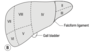
Relations of the spleen
Anteriorly: stomach
Posteriorly: left diaphragm, separating it from left pleura, lung, ribs 9, 10, 11
Inferiorly: splenic flexure of colon
Medially: left kidney
Route of the sperm (+ vas deferens anatomy)
Semineferous ducts in the lobules of the testis
Rete testes
Vasa in the epididymis
Into the vas deferens: 45cm, thick muscular tube
Through the scrotum - inguinal canal - lateral wall of the pelvis below the peritoneum - towards the ischial spine - turns medially towards base of bladder
Unites with seminal vesicles to form the common ejaculatory ducts
Enter bladder at the most superior and posterior aspect
Traverses bladder and opens in prostatic urethra at the verumonatnum - either side of the utricle

Capsules of the prostate
True capsule: thin, fibrous sheath surrounding the prostate
False capsule: condensation of the extraperioenal fascia, continuous with the fascia of the bladder and fascia of denonvilliers
Third capsule: sometimes created in BPH due to condensation of the periopheral part of the prostate gland
In enucleation of the prostate for BPH- the plane between the adenomatous mass and third capsule is enteredbetween te
Where is the venous plexus of the prostate?
In between the true and false fascia
True: thin fibrous capsule
False: condensation of the extraperitoneal fascia - continuous wiht the fascia around the bladder, and the fascia of denonvilliers posteriorly
Contents of the spermatic cord
3 layers of fascia
external spermatic (from ext oblique apo)
cremasteric (from int oblique apo)
internal spermatic (from transversalis fascia)
3 arteries
testicular
cremasteric
artery to the vas
3 nerves
sympathetic
ilioinguinal (lies on the cord)
genital branch of the genitofemoral N (to cremaster)
3 other things
vas deferens
pampiniform plexus of the veins
lymphatics
Main tributaries of the IVC
Lumbar branches
Right gonadal vein
Right renal vein
Left renal vein
Right suprarenal vein
Phrenic vein
Hepatic vein
Internal oblique
Origin
Insertion
Direction of fibres
Anterior 2/3 iliac crest, lumbodorsal fascia, lateral 2/3 inguinal ligament
linea alba, ribs 11/12, pubic crest via conjoint tendon
Upwards and medially
Parts of the penis
Root
Body
Glans
Transverse colon features
Relations: anterior, posterior, superior, inferior
Covered in mesentery - transverse mesocolon attached to the anterior surface of the pancreas
Becomes descending colon at the splenic flexure
Superiorly: liver, gallbladder, greater curvature of stomach, spleen
Inferiorly: coils of small intestine
Anteriorly: greater omentum
Posteriorly: right kidney, small intestine, left kidney, second part of duodenum, pancreas
Devleopment abnormalities in the kidney
Metanephric duct structures fails to fuse with metanephros structures = ARPKD
Metanephric duct may branch early - extra ureters; may extend into urethra/vagina - causing incontinence
Metanephros fails to develop one side - congenital absence of kidney
Two metanephric masses may fuse = horseshoe kidney
Kidney fail to migrate = pelvic kidney
Distal arteries may persist = aberrant renal arteries,

Subcostal (Kocher’s)
Indications
Process
Structures encountered
Risks
Cholecystectomy (right) / elective splenectomy (left) / Anterior approach for kidneys (both connected in middle)
2.5cm below and parallel to costal margin, extending laterally to border of rectus sheath or further
Skin, subcut fat, campers, scarpas, anterior rectus sheath, rectus abdominis, posterior rectus sheath, extraperitoneal fat, peritoneum
9th intercostal nerve near the lateral border of incision - if damaged - weakness and atrophy of upper rectus - predisposing to incisional hernia
Iliacus
Origin
Insertion
Nerve supply
Action
Greater part of iliac fossa extending onto sacrum
Lateral part of psoas major tendon onto the lesser trochanter
branch of femoral nerve (L2-L3)
Flexion of vetebral column
Rectus sheath:
above costal margin
above arcuate line
below arcuate line
Fuses in the middle = linea alba - xiphisternum - pubic symphysis
Anterior: External oblique only
Anterior: external oblique + 1/2 internal oblique
Posterior: transversus abdominis + 1/2 internal oblique; transversalis fascia, peritoneum
Anterior: external oblique + internal oblique + transversus; transversalis fascia + peritoneum
Distinguishing between jejunum and ileum
Jejunum is thicker as valvulae coniventes / plicae circulares are larger and more numerous
Greater diameter
Mesentery at jejunum - less arcades that are longer and straigher (less arches), less fatty and thinner vs lower down
Found at or above level of the umbilicus, whilst ileum more likely below
Quadratus lumborum
Origin
Insertion
Nerve supply
Action
Iliolumbar ligament and adjacent portion of iliac crest
Into the lower border of 12th rib medially and tendons into L1-L4 transverse processes
T12-L4
Extension and lateral flesion of lumbar vertebrae; fixes 12th rib during inspiration

Nerve supply of the oesophagus
Upper third
Below the root of the lung
Upper third: parasympathetic via RLN + sympathetic via middle cervical ganglion along inferior thyroid artery
Below root of the lung: Vagus + sympathetic nerves form the oesophageal plexus
Stomach
Anatomy: shape, surfaces, curves, orifices
Junction of body and pyloric antrum is called?
Pylorus with duodenum junction?
J shape, anterior and posterior surface, greater and lesser curvature, cardia and pylorus orifice
Cardia, body (with fundus at the top - lies above cardia), pylorus (antrum and canal)
Incisura angularis
constant prepyloric veins of Mayo - crosses it vertically

Location of the deep inguinal ring
Important structure next to it
1cm above the midpoint of the inguinal ligament
Defect in the transversalis fascia
Located laterally to the inferior epigastric vessels
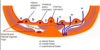
Blood supply of the spleen
Splenic artery from coeliac trunk (runs along upper border of pancreas)
Splenic vein - behind the pancreas
Splenic vessels + tail of the pancreas in the lieno-renal ligament + lymph nodes
Composition of the perineum
Anterior triangle (urogenital)
Posterior triangle (anal)
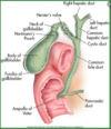
Relations of the pancreas
- anterior / posterior
Splenic vessels
The stomach and D1 lie in front of the pancreas - separated by mesentery (pancreas is retroperitoneal)
Transverse mesocolon attached to the anterior aspect of the pancreas
Below this is the DJ flexure, splenic flexure, small intestine
Posteriorly in contact with the left crus of the driaphragm, the aorta, left suprarenal gland and left kidney
Splenic artery = along upper border of pancreas
Splenic vein = being pancreas
Main duct from tail to head; accessory duct opens 2cm proximally in duodenum
Sites of narrowing of oesophagus
Commencement - 17cm from upper incisors
Where its crossed by left main bronchus - 28cm from upper incisors
Termination - 43cm from upper incisors