Anatomy Flashcards
Name proximal and distal attachment, action, and innervation of
STERNOCLEIDOMASTOID muscle
proximal attachment: mastoid process
**distal attachment: **manubrium of sternum, clavicle
**action: **bilateral contraction: flex cervical spine, unilateral contraction: laterally flex and rotate cervical spine
**innervation: **CN XI
Name proximal and distal attachment, action, and innervation of
ANTERIOR ABDOMINAL WALL muscles
proximal attachment: linea alba
**distal attachment: **thoracolumbar fascia
**action: **bilateral contraction: flex spine, unilateral contraction: laterally flex and rotate spine
**innervation: **thoracic ventral rami of spinal nerves
Name proximal and distal attachment, action, and innervation of
PSOAS muscle
proximal attachment: T12-L5 vertebral bodies and intervertebral disks
distal attachment: lesser trochanter of femur
action: flex spine (when attachment to femur is fixed)
innervation: L1-3 ventral rami of spinal nerves

Name proximal and distal attachment, action, and innervation of
QUADRATUS LUMBORUM muscle
proximal attachment: L5 vertebra, iliac crest
distal attachment: L1-L4 vertebrae, rib 12
action: bilateral contraction: extend spine, unilateral contraction: laterally flex spine
innervation: T12-L4 ventral rami of spinal nerves

Name proximal and distal attachment, action, and innervation of
SPLENIUS muscle
proximal attachment: C7-T3 spinous processes
distal attachment: mastoid process, occipital bone, C1-3 vertebrae
action: bilateral contraction: extend cervical and thoracic spine, unilateral contraction: laterally flex and rotate cervical spine
innervation: dorsal rami of spinal nerves

Name proximal and distal attachment, action, and innervation of
ERECTOR SPINAE muscles
proximal attachment: sacrum, spinous processes of lower vertebrae, iliac crest
distal attachment: 3 columns of muscles (iliocostalis, longissimus, spinalis) that insert on neural arches of vertebrae to the occiput
action: bilateral contraction: extend spine, unilateral contraction: laterally flex spine with some rotation
innervation: dorsal rami of spinal nerves

Name proximal and distal attachment, action, and innervation of
TRANSVERSOSPINAL muscles
proximal attachment: generalized C4-T12 transverse processes
distal attachment:
- semispinalis: extends to spinous processes across 4-6 spinal segments
- multifidus: extends to spinous processes across 2-4 spinal segments
- rotators: extend to spinous processes of adjacent spinal segments
action: extension and rotation of spine
innervation: dorsal rami of spinal nerves

Name proximal and distal attachment, action, and innervation of
SCALENE muscles
proximal attachment:
- anterior: C3-C6 transverse processes
- middle and posterior: C5-C7 transverse processes
distal attachment:
- anterior and middle: 1st rib
- posterior: 2nd rib
action: bilateral: flex cervical spine, unilateral: laterally rotate cervical spine
innervation: C3-C7 ventral rami of spinal nerves

Name proximal and distal attachment, action, and innervation of
LONGUS COLLI muscles
proximal attachment: C1-C6 vertebral bodies, transverse processes, and occiput
distal attachment: C3-T3 vertebral bodies and transverse processes
action: flex cervical spine
innervation: C1-C6 ventral rami of spinal nerves

Name proximal and distal attachment, action, and innervation of
PECTORALIS MAJOR muscle
proximal attachment: sternum, costal cartilages 1-6, clavicle
distal attachment: intertubercular (bicipital) groove of humerus
action: adducts and medially rotates the humerus; clavicular head (acting alone): flexes humerus; sternocostal head (acting alone): extends humerus from flexed position
innervation: lateral pectoral nerve, medial pectoral nerve

Name proximal and distal attachment, action, and innervation of
PECTORALIS MINOR muscle
proximal attachment: ribs 3-5
distal attachment: coracoid process of scapula
action: stabilizes and protracts scapula
innervation: medial pectoral nerve

Name proximal and distal attachment, action, and innervation of
SERRATUS ANTERIOR muscle
proximal attachment: ribs 1-9, outer surface
distal attachment: medial border of scapula
action: protracts scapula, raises ribs when scapula is fixed, stabilizes scapula
innervation: long thoracic nerve (C5-C7)

Name proximal and distal attachment, action, and innervation of
TERES MINOR muscle
proximal attachment: lateral border of scapula
distal attachment: greater tubercle of humerus
action: laterally rotates arm
innervation: axillary nerve

Name proximal and distal attachment, action, and innervation of
LATISSIMUS DORSI muscle
proximal attachment: posterior part of iliac crest, thoracolumbar fascia, T6-T12 spinous processes, ribs 3-4
distal attachment: intertubercular (bicipital) groove of humerus
action: at scapulothoracic joint: depress scapula, at shoulder joint: adduct, internally rotate (humerus), extend
innervation: thoracodorsal nerve
*partially overlapped by trapezius muscle

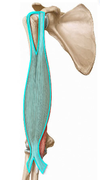
Name proximal and distal attachment, action, and innervation of
TRICEPS BRACHII muscle
proximal attachment:
- long head: infraglenoid tubercle of scapula
- lateral head: upper half of posterior shaft of humerus above spiral groove
- medial head: lower half of posterior shaft of humerus below spiral groove
distal attachment: olecranon process of ulna
action: at shoulder joint: extend (long head), at elbow joint: extend
innervation: radial nerve

Name proximal and distal attachment, action, and innervation of
BICEPS BRACHII muscle
proximal attachment:
- long head: supraglenoid tubercle of scapula
- short head: coracoid process of scapula
distal attachment: radial tuberosity, bicipital aponeurosis
action: at elbow joint: flex, at shoulder joint: flex (long joint), at radioulnar joint: supinate
innervation: musculocutaneous nerve
Name proximal and distal attachment, action, and innervation of
CORACOBRACHIALIS muscle
proximal attachment: coracoid process of scapula
distal attachment: medial shaft of humerus
action: at shoulder joint: flex, adduct
innervation: musculocutaneous nerve

Name proximal and distal attachment, action, and innervation of
PRONATOR TERES muscle
proximal attachment: medial epicondyle of humerus, coronoid process of ulna
distal attachment: lateral surface of radius
action: at radioulnar joints: pronate
innervation: median nerve

Name proximal and distal attachment, action, and innervation of
FLEXOR CARPI RADIALIS muscle
proximal attachment: medial epicondyle of humerus
distal attachment: base of 2nd and 3rd metacarpal
action: at wrist joint: flex, abduct
innervation: median nerve

Name proximal and distal attachment, action, and innervation of
PALMARIS LONGUS muscle
proximal attachment: medial epicondyle of humerus
distal attachment: flexor retinaculum and palmar aponeurosis
action: at wrist joint: flex (weak), tense palmar fascia
innervation: median nerve

Name proximal and distal attachment, action, and innervation of
FLEXOR CARPI ULNARIS muscle
proximal attachment: humeral head: medial epicondyle of humerus, ulnar head: olecranon process of ulna
distal attachment: pisiform bone; by ligaments to hook of the hamate and 5th metacarpal bone
action: at wrist joint: flex, adduct
innervation: ulnar nerve

Name proximal and distal attachment, action, and innervation of
EXTENSOR CARPI RADIALIS BREVIS muscle
proximal attachment: lateral epicondyle of humerus
distal attachment: base of 2nd and 3rd metacarpals (dorsal side)
action: at wrist joint: extend
innervation: radial nerve

Name proximal and distal attachment, action, and innervation of
EXTENSOR DIGITORUM muscle
proximal attachment: lateral epicondyle of humerus
distal attachment: extensor hood of fingers
action: at MCP joints of fingers: extend (NOTE: extend DIP/PIP joints through extensor hood)
innervation: radial nerve

Name proximal and distal attachment, action, and innervation of
EXTENSOR DIGITI MINIMI muscle
proximal attachment: lateral epicondyle of humerus
distal attachment: extensor hood of little finger
action: at MCP joint of little finger: extend (NOTE: extend DIP/PIP joints of little finger through extensor hood)
innervation: radial nerve

Name proximal and distal attachment, action, and innervation of
EXTENSOR CARPI ULNARIS muscle
proximal attachment: lateral epicondyle of humerus
distal attachment: base of 5th metacarpal (dorsal side)
action: at wrist joint: extend, adduct
innervation: radial nerve

Name origin and insertion, action, and innervation of
TRAPEZIUS muscle
origin: occipital bone, ligamentum nuchae, C7-T12 vertebrae
insertion: clavicle, acromion and spine of scapula
action:
- superior fibers elevate scapula
- middle fibers retract scapula
- inferior fibers depress scapulae
- superior and inferior fibers together cause upward or superior rotation of scapula
innervation: CN XI

Name origin and insertion, action, and innervation of
RHOMBOIDEUS MAJOR/MINOR muscle
origin: ligamentum nuchae, C7-T5 vertebrae
insertion: scapula - medial border (from spine to inferior angle)
action: retracts, inferiorly rotates, and stabilizes scapula
innervation: dorsal scapular nerve
*deep to trapezius muscle, inferior to levator scapulae

Name origin and insertion, action, and innervation of
LEVATOR SCAPULAE muscle
origin: C1-C4 vertebrae
insertion: scapula - superomedial border
action: elevates and inferiorly rotates scapula
innervation: dorsal scapular nerve
*deep to trapezius muscle, superior to rhomboideus major/minor

SUPERFICIAL BACK MUSCLES
Considered extrinsic “back” muscle (extrinsic muscles of the shoulder - posterior group). Connect the upper limbs to the trunk and control limb mvts. Innervated by ventral rami nerves (primarily from the brachial plexus).
- trapezius
- latissimus dorsi
- rhomboideus major/minor
- levator scapulae
INTERMEDIATE BACK MUSCLES
Considered extrinsic “back” muscles, function as accessory respiratory muscles. Innervated by ventral rami nerves.
- serratus posterior superior
- serratus posterior inferior
DEEP or TRUE BACK MUSCLES
Considered the intrinsic muscles of the back (act on vertebral column). Lie deep to extrinsic muscles and arranged into 3 layers: superficial, intermediate, and deep. Innervated by dorsal rami, collectively called the “paraspinals” clinically.
- superficial: splenius (capitis, cervicis)
- intermediate: erector spinae (iliocostalis, longissimus, spinalis)
- deep: transversospinalis (semispinalis - capitis, cervicis; multifidus; rotatores; interspinous and intertransverse muscles)
THORACOLUMBAR FASCIA
- dense connective tissue that surrounds and covers the deep (intrinsic) back muscles
- posterior aspect covers superficial layer of intrinsic back muscles
Types and number of vertebrae
- 7 cervical
- 12 thoracic
- 5 lumbar
- 5 sacral (fused into 1 bone)
- 4 coccygeal
- TOTAL: 33 bones
Curvatures of the vertebral column
- primary: present at birth (concave opening anteriorly), persist in thoracic and sacrococcygeal regions
- secondary: develop after birth in cervical and lumbar region

Movements of the vertebral column
- flexion: greatest in cervical region
- extension: greatest in lumbar region
- lateral bending: greatest in lumbar region
- rotation: greatest in thoracic region

Label
Parts of typical vertebra


Cervical, thoracic, and lumbar vertebra differences
- cervical: have bifid spinous process, transverse foramen (for vertebral artery)
- thoracic: have costal and demifacets (for ribs) and inferiorly oriented spinous processes
- lumbar: lack costal facets, have quadrangular and horizontally oriented spinous processes

Atypical vertebrae - Atlas, Axis
- atlas (C1): articulates superiorly with occipital bone, NO body or spinous process (instead has anterior and posterior arch, facet for dens). Atlanto-occipital joint allows for head flexion and extension.
- axis (C2): has an odontoid process (dens) around which the atlas rotates. Median atlanto-axial joint allows for rotation of head.
Atypical vertebrae - Sacrum
Fusion of S1-S5 vertebrae. Consists of body of S1, ala, promontory, anterior sacral foramina (transmits ventral rami of S1-S5 spinal nerves), posterior sacral foramina (dorsal rami of S1-S5), sacral canal, sacral hiatus*
***Caudal anesthesia may be administered through the sacral hiatus. The solutions pass superiorly in the loose connective tissue and bathe the spinal nerves as they emerge from the dural sheath. OBs use this method of nerve block to relieve labor pain (1st and 2nd stage). This does not affec the infant.
Locating the L4 spinous process
A horizontal line drawn across the iliac crests will intersect the L4 spinous process.
***important landmark in doing lumbar punctures to sample CSF
Zygapophyseal (facet) joints
- Synovial joints formed between articular processes of adjacent vertebrae
- permit gliding movements between vertebrae
- stabilized by ligaments uniting laminae, transverse and spinous processes
- innervated by dorsal rami nerves
Intervertebral discs
- Cartilaginous joints (specifically symphyses) between adjacent vertebral bodies designed for weight bearing and strength
- Composed of outer, tough anulus fibrosus (strength) and inner, gelatinous nucleus pulposus (shock absorption during weight bearing)
- ***major role in development of curvatures of vertebral column
Synovial joints
- “diarthroses” - highly moveable joints
- consist of: fibrous joint capsule (encloses joint cavity), synovial membrane (lines inside of fibrous capsule), synovial fluid (secreted by synovial membrane), hyaline cartilage (caps the ends of articulating bones)

Anatomical classification of joints
- Cartilaginous joints - symphyses, synchondroses
- Fibrous joints - syndesmoses, gomphoses, sutures
- Synovial joints - hinge type, pivot type, condyloid, saddle type, ball and socket, gliding (plane type)
Functional classification of joints
- Immoveable joints (synarthroses) - sutures, gomphoses, synchondroses, schindylesis
- Slightly moveable joints (amphiarthroses) - symphysis, syndesmoses
- Highly moveable joints (diarthroses) - hinge type, pivot type, condyloid, saddle type, ball and socket, gliding
Cartilaginous joints
- formed by 2 bones separated by cartilage of some type
- 2 types: symphyses (midline of body, e.g. intervertebral discs), synchondroses
Intervertebral (neural) foramen
formed when 2 vertebrae come together
transmits spinal nerves
Vertebral (spinal) canal
- formed by articulation of vertebral foramen of adjacent vertebrae
- transmits spinal cord
Label and name the function of
Vertebral column ligaments

- Anterior longitudinal ligament: prevents hyperextension of vertebral column
- Posterior longitudinal ligament: prevents hyperflexion
- Supraspinous and interspinous ligaments: stabilize vertebral column
- Ligamentum flavum: unites laminae of adjacent vertebrae, elastic nature preserves curvatures of vertebral column

Meninges
- Formed by 3 membranes (superficial to deep): dura mater, arachnoid mater, pia mater
- Covers spinal cord
Meningeal spaces
- epidural space: space superficial to dura mater
- subdural space: space between dura mater and arachnoid mater
- subarachnoid space: space between arachnoid mater and pia mater, contains CSF that cushions spinal cord
- ***hematomas can occur in subdural and epidural spaces
Denticulate ligament
- Serrated, tooth-like ligament extending between the dorsal and ventral roots
- Formed by pia mater
- Helps anchor spinal cord within dural sac
Spinal cord segmentation
A spinal cord segment is a section of spinal cord that gives rise to a pair of spinal nerves (dorsal and ventral rami nerves) on each side. A dermatome is a strip of skin that is innervated by sensory fibers from a single spinal cord segment.
- 8 cervical
- 12 thoracic
- 5 lumbar
- 5 sacral
- 1 coccygeal
Dorsal and ventral roots of spinal nerves
Sensory fibers coming into the spinal cord form a dorsal root. Along the dorsal root is a dorsal root ganglion (DRG) which contains sensory cell bodies.
Motor fibers leaving the spinal cord form a ventral root (the cell bodies for these motor fibers are within the ventral horn of gray matter within the spinal cord).
Spinal nerves
There are 31 pairs of spinal nerves. A dorsal and ventral root fuse to form a short spinal nerve.
Dorsal and ventral rami nerves
Each spinal nerve quickly divides into two main branches (branches = “rami” in latin): a dorsal ramus nerve and a ventral ramus nerve.
The dorsal rami nerves innervate the intrinsic or deep/true back muscles on the posterior 1/3 of the body wall (soma) circumference.
The ventral rami nerves innervate the antero-lateral 2/3rds of the body wall circumference.
Spinal cord vs. Vertebral column length
The spinal cord is MUCH SHORTER than the vertebral column. The spinal cord ends opposite L2.
Nerve roots and vertebrae relationship
In the cervical region the spinal nerves emerge (in the intervertebral foramen) ABOVE their corresponding vertebra.
However, because there are only 7 cervical vertebra, the C8 spinal nerve emerges BELOW the C7 vertebra.
From T1 down, the spinal nerves emerge BELOW their corresponding vertebra.
Filum terminale
ligament-like structure that eventually anchors to coccyx along with dura and arachnoid mater, forming the coccygeal ligament
helps provide stability for the spinal cord
***dural sac and subarachnoid space end opposite S2
Conus medullaris and cauda equina
As the spinal cord ends, it tapers down to form a cone-like structure called the conus medullaris (which ends at L2 vertebral level).
Surrounding it are dorsal and ventral roots that are proceeding inferiorly. These roots look like a “horse’s tail” and so we call it the cauda equina.
Extending from the conus medullaris and traveling through the cauda equina is the filum terminale.
***Remember, the dural sac (and subarachnoid space with CSF) ends at the S2 vertebral level, but the filum terminale continues inferiorly to anchor to the coccyx.
Blood supply of the spinal cord
- 1 anterior spinal artery
- 2 posterior spinal arteries (from vertebral arteries)
- radicular arteries from segmental spinal arteries (from posterior intercostal arteries)
- drained by 3 anterior and 3 posterior spinal veins -> form internal vertebral venous plexus (Batson’s plexus)
Internal vertebral venous plexus (Batson’s plexus)
formed by veins draining spinal cord and vertebrae
network of valveless veins that is continuous with cranial dural venous sinuses within skull -> provides pathway for infection between head and lower parts of body!
CNS
brain - 12 pairs of cranial nerves
spinal cord - 31 pairs of spinal nerves
Identify
Osteology: anterior view


Identify
Osteology: lateral view


Identify
Osteology: lateral view


Cutaneous innervation of the face
- V1 (ophthalmic): supraorbital nerve
- also supratrochlear, infratrochlear, lacrimal, external nasal
- V2 (maxillary): infraorbital nerve
- also zygomaticotemporal, zygomaticofacial
- V3 (mandibular): mental nerve
- also auriculotemporal, buccal
Cutaneous innervation of the scalp
- V1: supraorbital nerve
- V2: zygomaticotemporal nerve
- V3: auriculotemporal nerve
- C2, C3: lesser occipital nerve
- C2: greater occipital nerve
***C1 has NO cutaneous branch to head and neck. Thus dermatomal pattern comprised only of V1-3, C2-4
Muscles of facial expression: superior and orbital group
Occipitofrontalis: frontalis part elevates eyebrows, occipitalis part tenses galea aponeurotica/epicranial aponeurosis
Orbicularis oculi: closes eyelids
Corrugator supercilli: draws eyebrows medial and downwards

Muscles of facial expression: nasal group
Procerus: draws down medial angle of eyebrows producing transverse wrinkles over bridge of nose
Nasalis: compresses nasal aperture and laterally opens nostrils

Muscles of facial expression: oral group
Buccinator: compression of the cheek, positioning food between teeth
Depressor anguli oris: draws corner of mouth inferiorly and laterally
Zygomaticus major: draws corner of mouth superiorly and laterally
Zygomaticus minor: draws upper lip upward
Orbicularis oris: closure of mouth, protrusion of lips
Mentalis: raises and protrudes lower lip as it wrinkles skin of chin
Risorius: retracts corner of mouth
Depressor labii inferioris: draws lower lip downward and laterally
(Platysma: depresses lower jaw, tenses skin of neck)

Facial nerve motor branches to muscles of facial expression
- temporal: to temporal region
- zygomatic: to zygomatic region
- buccal: to cheek or buccal region
- mandibular: to margin of mandible area
- cervical: to anterior neck region
Functional components of nerves - Somatic afferent
- somatic sensory fibers from skin, striated skeletal muscle, joints of the soma
- convey info from pain, temp, touch, proprioception
Functional components of nerves - Somatic efferent
- somatic motor fibers to striated skeletal muscle of the soma
Functional components of nerves - Visceral afferent
- visceral sensory fibers from glands, blood vessels, smooth muscle, cardiac muscle
Functional components of nerves - Visceral efferent
- visceral motor fibers to glands, smooth muscle, cardiac muscle
- aka autonomic nervous system
Functional components of nerves - Special sense
- sensory fibers from the 5 senses: nose (smelling), eyes (sight), ears (hearing and balance), tongue (taste)
Functional components of nerves - Branchial motor
- motor fibers to striated, skeletal muscle of the head and neck which has developed embryologically from the branchial/pharyngeal arches (NOT from somites)
- muscles innervated include muscles of facial expression, mastication, and pharynx/larynx
*
Name the cranial nerve, muscle, and cartilage/bony derivatives of
Branchial arch 1
- CN V (trigeminal)
- muscles of mastication, mylohyoid, anterior belly of digastric, tensor vili palatini, tensor tympani
- malleus, incus, mandible
Name the cranial nerve, muscle, and cartilage/bony derivatives of
Branchial arch 2
- CN VII (facial)
- muscles of facial expression, posterior belly of digastric, stylohyoid, stapedius
- stapes, styloid, part of hyoid
Name the cranial nerve, muscle, and cartilage/bony derivatives of
Branchial arch 3
- CN IX (glossopharyngeal)
- stylopharyngeus
- part of hyoid
Name the cranial nerve, muscle, and cartilage/bony derivatives of
Branchial arches 4 & 6
- CN X (vagus)
- muscles of pharynx, larynx, palate
- larynx
Axial skeleton
bones of:
the head (skull)
neck (hyoid bone and cervical vertebrae)
trunk (ribs, sternum, vertebrae, sacrum)
Appendicular skeleton
bones of the limbs (extremities, appendages), including those forming the pectoral (shoulder) and pelvic girdles
Glenohumeral joint
The tendon of which muscle passes through this joint?
- synovial joint (ball and socket)
- allows for flexion, extension, abduction, adduction, medial and lateral rotation, circumduction of humerus
- innervated by: suprascapular, lateral pectoral, axillary nerves
- **high mobility of this joint results in poor stability and joint is often dislocated
The tendon of the long head of the biceps muscle passes through this joint. It is held in the bicipital groove by the transverse humeral ligament.
Foramen of Weitbrecht
weakness in joint capsule between superior and middle glenohumeral ligaments
**head of the humerus penetrates through this weak area in an anterior dislocation of the shoulder!
How is the glenohumeral joint reinforced?
reinforced by rotator cuff tendons, anteriorly by glenohumeral ligaments (thickenings of the joint capsule)
The tendon of the long head of the biceps invaginates the joint capsule but does not enter the synovial cavity (intracapsular and extrasynovial)
Glenoid labrum
fibrocartilagenous ring that surrounds the glenoid fossa and helps deepen the socket of the shoulder joint
**the fibrous joint capsule attaches directly to the labrum, so any injury of the joint capsule can potentially involve the labrum!
Sternoclavicular (SC) joint
- synovial joint (saddle-type, but functions like a ball and socket)
- joint separated into 2 separate joint cavities by an articular disk
- very mobile to allow movements of the pectoral girdle (clavicle + scapula)
- it is the ONLY joint between the pectoral girdle and axial skeleton
- innervated by: supraclavicular nerves, nerve to subclavius muscle
- **very strong joint -> dislocation is rare
Acromioclavicular (AC) joint
- synovial joint (plane type - allows gliding movements)
- protected by strong superior and inferior acromioclavicular ligaments (also coracoacromial and coracoclavicular ligaments)
- innervated by: supraclavicular, lateral pectoral, and axillary nerves
- **this is the joint involved in shoulder separation injuries!
Suprahumeral joint
- not a true joint! refers to the space between the head of the humerus and the acromion and coracoacromial ligament
- space contains: biceps long head tendon, rotator cuff tendons, subacromial/subdeltoid bursa, glenohumeral joint capsule
- **thus inflammation of suprahumeral joint can impinge these structures -> possible frozen shoulder syndrome
Extrinsic muscles of the shoulder - anterior group
- pectoralis major
- pectoralis minor
- subclavius
- serratus anterior
Extrinsic muscles of the shoulder - posterior group
same as superficial “back” muscles:
- trapezius
- levator scapulae
- rhomboids
- latissimus dorsi
Movements of the scapula and muscles responsible
- elevation: trapezius (superior fibers), levator scapulae
- depression: trapezius (inferior fibers)
- retraction: trapezius (middle fibers), rhomboids
- protraction: serratus anterior
- superior (upward) rotation: trapezius (superior and inferior fibers), serratus anterior
- inferior (downward) rotation: levator scapulae, rhomboids
Name proximal and distal attachment, action, and innervation of
SUBCLAVIUS muscle
proximal attachment: 1st rib and its costal cartilage
distal attachment: clavicle (inferior surface)
**action: **draws clavicle inferiorly and anteriorly, protects structures deep to clavicle
**innervation: **nerve to subclavius
Scapulohumeral rhythm
integrated movement of the scapula and humerus together
- involved in movement of upper limb to place hand in functional position
- for every 2 degrees of abduction at the glenohumeral joint, there is 1 degree of scapular rotation!
- e.g. abduction of arm 90 degrees - requires simultaneous movement of humerus at glenohumeral joint (60 degrees) accompanied by rotation of scapula (30 degrees).
Intrinsic muscles of the shoulder
- deltoid
- teres major
- supraspinatus
- infraspinatus
- teres minor
- subscapularis
Name proximal and distal attachment, action, and innervation of
SUPRASPINATUS muscle
proximal attachment: scapula (supraspinous fossa)
**distal attachment: **humerus (greater tubercle)
**action: **initiates abduction, laterally (externally) rotates humerus
**innervation: **suprascapular nerve
Name proximal and distal attachment, action, and innervation of
INFRASPINATUS muscle
proximal attachment: scapula (infraspinous fossa)
**distal attachment: **humerus (greater tubercle)
**action: **laterally rotates arm
**innervation: **suprascapular nerve
Rotator cuff
- the cuff is actually the conjoined tendon of the 4 rotator cuff muscles around the fibrous joint capsule of the humerus
- function: reinforces shoulder joint on all sides (exc inferiorly -> thus weak here)
Critical zone (of the rotator cuff)
- area of vascular anastomoses within the rotator cuff
- alternately ischemic or hyperemic depending on whether there is tension on the cuff
- **often the site of degeneration, Ca++ deposits, and tears
Scapular anastomosis
- consists of:
- branches of the 1st part of the subclavian artery: transverse cervical artery, suprascapular artery, dorsal scapular artery
- branches of the 3rd part of the axillary artery: subscapular artery and its branches
- intercostal arteries
- demonstrates principle of collateral circulation - permits blood to bypass occlusion and still get to its destination
A fracture of the surgical “neck” of the humerus will endanger what structures coursing through the quadrangular space?
posterior circumflex humeral artery
axillary nerve
(quadrangular space bordered by triceps lateral head and long head, teres major and minor muscles)
Brachial plexus
somatic plexus formed by ventral rami of C5-T1 spinal nerves (or spinal cord segments)
provides sensory and motor innervation to upper limb (pectoral region, shoulder region, arm, forearm, hand) and all pectoral girdle muscles, except the trapezius
divided into 2 parts: supraclavicular (in posterior neck), infraclavicular (in axilla)
consists of 5 roots, 3 trunks, 6 divisions, 3 cords, 5 terminal branches
ID the five regions of the brachial plexus and their components
5 roots: C5-T1 ventral rami
3 trunks: upper (superior), middle, lower (inferior)
6 divisions: anterior (innervate flexor muscles via musculocutaneous, median, ulnar nerves) and posterior (innervate extensor muscles via radial nerve)
3 cords: lateral, posterior, medial
5 terminal branches: axillary, radial, musculocutaneous, ulnar, median nerves
5 terminal branches of the brachial plexus, the spinal cord segments they represent, and their motor innervation
axillary nerve (C5-C6): deltoid, teres minor
musculocutaneous nerve (C5-C7): muscles of the anterior (flexor) compartment of the arm - biceps, brachialis, coracobrachialis
median nerve (C5-T1): muscles of the anterior (flexor) compartment of the forearm (exc 1 1/2 muscles) and muscles in 1 1/2 compartments of the hand [pronate forearm, flex wrist and fingers, move thumb]
ulnar nerve (C8-T1): 1 1/2 muscles in flexor compartment of forearm and most of the muscles in the hand [flex wrist and move fingers]
radial nerve (C5-T1): muscles of the posterior (extensor) compartment of arm and forearm [extension at the shoulder, elbow, wrist]
Non-terminal branches of the brachial plexus and their innervation
-
roots:
- long thoracic nerve - serratus anterior
- dorsal scapular nerve - levator scapulae, rhomboids
-
upper trunk:
- suprascapular nerve - supraspinatus, infraspinatus
- nerve to subclavius - subclavius
-
medial cord:
- medial pectoral nerve - pec. major and minor
- medial brachial cutaneous nerve - sensory to medial side of arm
- medial antebrachial cutaneous nerve - sensory to medial side of forearm
-
lateral cord:
- lateral pectoral nerve - pec. major
-
posterior cord:
- upper subscapular nerve - subscapularis
- thoracodorsal nerve - lat. dorsi
- lower subscapular nerve - subscapularis, teres major
Boundaries of the axilla
- apex
- medial wall - lateral bony thorax, serratus anterior
- anterior wall - pec major and minor, subclavius, clavicle
- lateral wall - bicipital (intertubercular) groove of humerus
- posterior wall - scapular, subscapularis, teres major, latissimus dorsi
- base - skin and superficial fascia
Contents of the axilla
- vessels: axillary artery and vein
- lymph nodes: axillary lymph nodes (lymph from upper limb and 75% from breast)
- muscles and tendons: biceps long head tendon, biceps short head tendon, coracobrachialis
- brachial plexus: infraclavicular portion (cords and terminal branches)
Axillary artery, parts, and branches
- originates from subclavian artery at 1st rib and ends at inferior border of teres major to become brachial artery
- divided into 3 parts by pec minor:
- medial - supreme thoracic artery
- deep - thoracoacromial artery, lateral thoracic artery
- lateral - anterior circumflex humeral artery, posterior circumflex humeral artery, subscapular artery
Boundaries of the posterior triangle of the neck
SCM (posterior portion), trapezius (anterior portion), and clavicle
Roof of the posterior triangle of the neck
- formed by the investing fascia of the cervical fascia
- contains cutaneous branches of the cervical plexus:
- transverse cervical nerve (C2, C3)
- great auricular nerve (C2, C3)
- lesser occipital nerve (C2)
- supraclavicular nerves (C3, C4)
Muscular floor of the posterior triangle of the neck
- splenius capitis
- levator scapulae
- posterior scalene
- middle scalene
- anterior scalene
Brachial plexus in posterior triangle of neck
supraclavicular brachial plexus (roots and trunks)
- between anterior and middle scalene muscles, superior to subclavian artery, courses over 1st rib
What nerve controlling respiration courses through the posterior triangle? What muscle does it control, and what muscle is it related to?
phrenic nerve (component of the cervical plexus - C1-C4 ventral rami somatic plexus)
- innervates the diaphragm
- courses inferiorly on anterior surface of anterior scalene
What artery is located immediately deep to the clavicle? What is its origin and destination?
suprascapular artery
- courses posteriorly across floor of posterior triangle, deep to clavicle, to shoulder region to supply supraspinatus and infraspinatus
- originates from thyrocervical trunk of subclavian artery (1st part), destination is supraspinatus and infraspinatus
What artery coursing through the posterior triangle is supplying the trapezius muscle?
transverse cervical artery
- originates from thyrocervical trunk of subclavian artery (1st part)
- relationship: courses posteriorly across floor of posterior triangle, superior to suprascapular artery to deep surface of trapezius (to supply it)
What organ would be in danger of being pierced by a knife directed inferiorly into the posterior triangle?
lungs
What divides the arm into anterior and posterior compartments? What is the clinical signficance of these kind of compartments?
Humerus, intermuscular fascial septum
Muscles in each compartment are responsible for one particular function??
What is the general function of the muscles in the anterior compartment of the arm?
Flexor compartment - contains 3 muscles (BBC) that flex the humerus at the shoulder and/or the elbow joints
What are the muscles of the anterior compartment of the arm? Which muscle only crosses the shoulder joint anteriorly? Which muscle only crosses the elbow joint anteriorly? Which muscle crosses both the elbow and shoulder joints anteriorly?
BBC
Coracobrachialis only crosses the shoulder joint anteriorly
Brachialis only crosses the elbow joint anteriorly
Biceps brachii crosses both the elbow and shoulder joints anteriorly
Which muscle of the anterior compartment of the arm is also a powerful supinator?
Biceps brachii
What nerve innervates all the muscles of the anterior compartment of the arm?
Musculocutaneous nerve
(The median and ulnar nerves travel through the anterior compartment in the medial neurovascular bundle, but they do NOT innervate anything in the arm. They will innervate the anterior compartment of the forearm and compartments of the hand.)
What is the artery to the anterior compartment of the arm?
Brachial artery - continuation of the axillary artery and travels down the arm in the medial neurovascular bundle. The brachial artery gives rise to the deep brachial artery (profunda brachii a.) as it enters into the arm. The deep brachial artery will travel into the posterior compartment of the arm and will accompany the radial nerve in the radial groove of the humerus deep to the triceps.
What are the contents of the medial neurovascular bundle of the anterior compartment of the arm? Is the musculocutaneous nerve contained within this neurovascular bundle?
brachial artery, brachial veins, basilic vein, median nerve, ulnar nerve, and medial antebrachial cutaneous nerve
The musculocutaneous nerve is NOT contained within this neurovascular bundle.
Which muscle of the anterior compartment of the arm is pierced through its belly by the musculocutaneous nerve?
Coracobrachialis - musculocutaneous nerve pierces through belly of coracobrachialis, and then sits between superficial and deep muscles of the anterior compartment of the arm
What are the main structures (muscle, nerve, artery) of the posterior compartment of the arm?
triceps (medial, lateral, and long heads), radial nerve, deep brachial artery
Which head of the triceps crosses the teres major muscle superficially?
Long head??
Which head of the triceps crosses the shoulder joint posteriorly?
Long head of the triceps
What is the relationship of the radial nerve and deep brachial artery to the triceps and humerus?
Radial nerve - innervates the triceps. It lies deep to the triceps and travels in the radial groove of the humerus along with the deep brachial artery.
Deep brachial artery (profunda brachii) - branch of the brachial artery and travels with the radial nerve in the radial groove.
What is the function of the triceps?
All heads are involved with extending forearm at the elbow joint
Long head extends arm at shoulder joint
What structures cross the anterior aspect of the cubital fossa superficially from lateral to medial? How would you locate the medial nerve in the cubital fossa to do a nerve block?
Lateral to medial:
- **lateral antebrachial cutaneous nerve: **continuation of the musculocutaneous nerve after it gives off its motor fibers to the anterior compartment muscles and then travels lateral to the biceps tendon.
- biceps tendon: attaches to both the radial tuberosity and the bicipital aponeurosis which passes obliquely over the brachial artery and median nerve and is continuous with the deep fascia of the forearm.
- brachial artery: runs in the medial neurovascular bundle of the arm and will divide in the proximal forearm into two terminal branches: the radial and ulnar arteries
- median nerve: runs in the medial neurovascular bundle of the arm. Along with the ulnar nerve, these two nerves just pass through the arm without innervating anything.
Medial nerve block?
Where would you find the radial nerve in the cubital fossa and what are its two terminal branches?
To find the radial nerve, look deep in between the brachialis and brachioradialis muscles.
The radial nerve divides into the deep radial nerve (innervates the supinator, then pierces it to get access to the posterior compartment of the forearm where it will innervate all of the muscles of that compartment) and superficial radial nerve (cutaneous nerve that travels deep to the brachioradialis)
What nerve travels in the medial neurovascular bundle and then goes posterior to the medial epicondyle of the humerus and then goes back into the anterior compartment of the forearm?
Ulnar nerve
The forearm is divided into compartments by what structures?
Fascial intermuscular septa, interosseous membrane between the radius and ulna
What is the general function of the anterior compartment of the forearm?
contains muscles which will flex the wrist and/or digits
“flexor” compartment
What are the muscles of the superficial layer (flexor-pronator group) in the anterior compartment?
pronator teres, flexor carpi radialis, palmaris longus, flexor carpi ulnaris
What is the muscle of the intermediate layer of the anterior compartment of the forearm?
flexor digitorum superficialis
What are the muscles of the deep layer of the anterior compartment of the forearm?
flexor digitorum profundus, flexor pollicis longus, pronator quadratus
What action, aside from flexion, does flexor carpi ulnaris and flexor carpi radialis do?
Flexor carpi ulnaris also does ADDUCTION of the hand (ulnar deviation).
Flexor carpi radialis also does ABDUCTION of the hand (radial deviation).
What is unusual about the palmaris longus muscle?
This muscle is variable. Some people have it and some people don’t. And some people may even have it on one side but not the other!
The flexor digitorum superficialis flexes primarily at what joints?
medial 4 digits at proximal IP joints
The flexor digitorum profundis flexes primarily at what joints?
medial 4 digits at distal IP joints (but can also be recruited for MP joints and wrist)
What does flexor pollicis longus do?
Flexes the thumb
Which muscles of the anterior compartment are innervated by the ulnar nerve? Which are innervated by the median nerve?
Ulnar nerve - Flexor carpi ulnaris, 1/2 of flexor digitorum profundus
Median nerve - all muscles of the anterior compartment except flexor carpi ulnaris and 1/2 of flexor digitorum profundus
What are the terminal branches of the brachial artery?
Radial and ulnar arteries
The anterior interosseus nerve is a branch of what nerve?
Median nerve
What is the origin of the common interosseus artery? And what branches does it give rise to? Where are these branches located?
The common interosseous artery is a branch of the ulnar artery. It gives rise to the anterior interosseous artery (anterior compartment) and posterior interosseous artery (posterior compartment).
What is the basic organization arrangement (in terms of layers and groups) of the musculature of the posterior forearm?
The muscles of the posterior compartment are arranged into superficial and deep layers; the superficial layer is further subdivided into a posterior and lateral group.
What is the general function of the muscles of the posterior compartment of the forearm?
extending at either the wrist, the digits, or both
What cutaneous vein travels up the medial side of the forearm? What cutaneous vein travels up the lateral side of the forearm?
The basilic vein travels up the medial side of the forearm.
The cephalic vein travels up the lateral side of the forearm.
In the superficial layer of muscles in the posterior compartment of the forearm, what muscles comprise the lateral group? Which muscles comprise the posterior group?
Lateral group:
- brachioradialis
- extensor carpi radialis longus
- extensor carpi radialis brevis
Posterior group:
- anconeus
- extensor carpi ulnaris
- extensor digitorum
- extensor digiti minimi
What main nerve, either directly or indirectly, controls the muscles and function of the posterior forearm compartment?
Posterior interosseous nerve, a branch of the radial nerve
What is the artery of the posterior compartment of the forearm?
Posterior interosseous artery (continuation of deep radial artery)
What are the muscles of the deep layer of the posterior forearm compartment?
- supinator
- abductor pollicis longus
- extensor pollicis brevis
- extensor pollicis longus
- extensor indicis
Which muscles of the posterior forearm compartment act on the digits (other than the thumb)?
- extensor digitorum
- extensor digiti minimi
- extensor indicis
Which muscles of the posterior forearm compartment act on the wrist?
- extensor carpi ulnaris
- extensor carpi radialis brevis
- extensor carpi radialis longus
Which muscle of the posterior forearm opposes the action of the pronator muscles of the anterior compartment?
Supinator
Which tendons form the immediate boundaries of the anatomical “snuff-box”?
- extensor pollicis brevis (along with abductor pollicis longus)
- extensor pollicis longus
- extensor retinaculum along the bottom
The pulse of what artery can be taken by palpating the floor of the snuff-box?
Radial artery
What carpal bone, if fractured, would be painful as you palpate the floor of the anatomical snuff-box?
Scaphoid
What are the extensor expansions?
flattening of extensor tendons distally (in digits)
What is the function of the extensor retinaculum?
band of deep fascia that keeps the extensor tendons in place when the extensor muscles contract
What bones are involved in the wrist joint?
- distal radius
- proximal carpal bones: scaphoid, lunate, triquetrum
What actions are allowed at the wrist?
flexion, extension, ulnar deviation (adduction), radial deviation (abduction), circumduction
What nerves innervate the wrist joint?
ulnar and median nerves?
From medial to lateral, what structures cross the wrist anteriorly?
Medial to lateral: flexor carpi ulnaris tendon, ulnar nerve, ulnar artery, palmaris longus tendon, median nerve, flexor carpi radialis tendon, radial artery
Which structure forms the roof of the carpal tunnel?
flexor retinaculum
Which structures are contained within the carpal tunnel?
- 4 flexor digitorum superficialis tendons
- 4 flexor digitorum profundus tendons
- flexor pollicis longus tendon
- median nerve
What would be the symptoms of inflammation and compression within the carpal tunnel? What nerve would be affected? How might those symptoms be relieved surgically?
Compression of the median nerve
- sensory: pain and sensory deficits (paresthesia, hypothesia, anesthesia) along the distribution of its cutanous branches, which innervate the 3 1/2 lateral digits of the hand
- motor: motor deficits to thenar compartment muscles and lateral 2 lumbrical muscles; muscle weakness, loss of coordination, inability to oppose the thumb
These symptoms may be relieved by carpal tunnel release, a partial or complete surgical division of the flexor retinaculum.
What are the four compartments of the hand?
- thenar
- hypothenar
- central (midpalmar)
- deep (adductor, interossei)
What muscles are contained within the thenar compartment and what are their functions?
3 muscles relating to the thumb:
- abductor pollicis brevis
- flexor pollicis brevis
- opponens pollicis
What muscles are contained within the hypothenar compartment and what are their functions?
3 muscles relating to the 5th digit:
- abductor digiti minimi
- flexor digiti minimi
- opponens digiti minimi
What structures are contained within the central compartment?
flexor tendons (FDS, FDP), 4 lubricals
What is the innervation pattern of the lumbrical muscles and what is their function?
Innervation pattern
- lateral 2: median nerve
- medial 2: ulnar nerve
Function: flex at the MCP joints and extend at the IP joints of digits 2-5
What muscles are contained within the deep compartment?
adductor pollicis, interossei (palmar and dorsal)
What is the function of the dorsal interossei muscles and how many are there?
4 dorsal interossei muscles; function to ABDUCT the digits
What is the function of the palmar interossei muscles and how many are there?
4 palmar interossei muscles; function to ADDUCT the digits
Describe the blood supply to the hand.
brachial artery -> ulnar and radial arteries in the forearm -> superficial and deep palmar arches -> common palmar digital arteries -> proper palmar digital arteries
Which artery is predominant in the formation of the superficial palmar arch?
Ulnar artery
Which artery is predominant in the formation of the deep palmar arch?
Radial artery
The deep palmar arch accompanies the deep branch of the ulnar nerve in a neurovascular plane between what two compartments?
Anterior and posterior compartments of the forearm?
Which compartment of the hand is completely innervated by the median nerve (in terms of its muscles)?
thenar compartment
Which muscles of the central compartment are innervated by the median nerve? Which are innervated by the ulnar nerve?
median nerve: lateral 2 lumbricals
ulnar nerve: medial 2 lumbricals
The muscles of the hypothenar compartment and deep compartment are innervated by what nerve?
Ulnar nerve
On the palmar aspect of the hand, which nerve supplies the skin of the lateral 3 ½ digits and related palm area?
Median nerve
On the palmar aspect of the hand, which nerve supplies the skin of the medial 1 ½ digits and related palm area?
Ulnar nerve
On the dorsum of the hand, which nerve supplies the skin of the lateral 3 ½ digits (up to the knuckles approximately) and related back of the hand area?
Superficial radial nerve
On the dorsum of the hand, which nerve supplies the skin of the tips of the lateral 3 ½ digits?
Median nerve
On the dorsum of the hand, which nerve supplies the skin of the medial 1 ½ digits and related back of the hand area?
Ulnar nerve
Why are dermatomal maps different from cutaneous nerve pattern maps? How might each be useful in the clinics?
A dermatome is a strip of skin innervated by fibers from a single spinal cord level. A cutaneous nerve contains fibers from several spinal cord levels.
Knowing these two different patterns allows one to test for peripheral nerve problems (cutaneous nerve innervation pattern) or neurologic (spinal cord or spinal nerve) level problems (dermatomal pattern).
What are the 3 articulations of the elbow joint?
humeroulnar, humeroradial, radioulnar
What nerves innervate the elbow joint?
Median and ulnar nerves?
At what articulations of the elbow joint does flexion/extension occur?
humeroulnar, humeroradial joints
At what articulation of the elbow joint does supination/pronation occur?
proximal radioulnar joint
What ligaments strengthen the elbow joint medially and laterally? How might the integrity of these ligaments be tested?
Radial collateral ligament (LCL), ulnar collateral ligament (MCL)
Integrity tested by supination/pronation?
What is the function of the annular ligament?
helps protect the proximal radioulnar joint and allows the head of the radius to move beneath it while preventing it from being displaced
Bursitis at the elbow would involve which bursae potentially?
Subtendinous olecranon bursa (deep to triceps tendon), subcutaneous olecranon bursa (in superficial fascia over olecranon process)
The arterial anastomosis around the elbow joint involves branches from which main arteries?
Brachial, deep brachial, ulnar, radial
What are the three gluteal muscles? What is their function and innervation?
Gluteus maximus
- action: main extensor of thigh, lateral rotator of thigh, helps steady the extended leg
- innerv: inferior gluteal nerve
Gluteus medius
- action: abducts and medially rotates thigh, helps steady the pelvis
- innerv: superior gluteal nerve
Gluteus minimus
- action: abducts and medially rotates thigh, helps steady the pelvis
- innerv: superior gluteal nerve
What is the “safe area” of the gluteal region for intramuscular injections?
Upper outer (lateral) quadrant; using any of the other three quadrants can put the sciatic nerve at risk and cause very serious damage
What is the function of the piriformis, gemelli, obturator internus, and quadratus femoris muscles?
laterally rotate extended thigh, all but quad. femoris also abduct flexed thigh
What spinal cord level is considered to be mainly responsible for extension at the hip?
L2-4 (femoral) ?
What is the relationship of the sciatic nerve to the piriformis muscle?
The sciatic nerve is inferior to the piriformis muscle.
The sciatic nerve is a branch of what nerve plexus?
Sacral plexus (largest branch, L4-S3)
The sciatic nerve innervates what compartments and regions of the lower limb?
tibial division of sciatic nerve: hamstrings in the posterior thigh
fibular division of sciatic nerve: short head of biceps femoris
tibial nerve: foot, posterior compartment of leg
fibular nerve: anterior and lateral compartments
What fascia completely surrounds the thigh like a “sock”?
fascia lata
What are the three hamstrings? What is their function (at the hip and the knee) and innervation?
semitendinosus
- action: extend hip and flex knee
- innervation: tibial division of sciatic nerve
semimembranosus
- action: extend hip and flex knee
- innervation: tibial division of sciatic nerve
biceps femoris
- action: flex knee and laterally rotate leg, long head - extend hip
- innervation: long head - tibial division of sciatic nerve, short head - common peroneal division of sciatic nerve
What is the common proximal attachment of the hamstring muscles?
ischial tuberosity (exc short head of biceps femoris - linea aspera of femur)
Which hamstring is innervated by both divisions of the sciatic nerve?
biceps femoris (long head - tibial division, short head - common peroneal division)
What are the boundaries of the popliteal fossa?
hamstrings and gastrocnemius muscles
What are the contents of the popliteal fossa?
popliteal artery and vein, tibial nerve, common peroneal nerve
What is the origin of the popliteal artery?
continuation of the femoral artery (after diving into adductor hiatus?)
Which nerve of the popliteal region will continue into the posterior compartment of the leg?
tibial nerve (innervates posterior compartment and foot. common peroneal nerve innervates anterior and lateral compartments of leg)
What compartments are controlled by the common peroneal (fibular) nerve? What are the general functions of these compartments?
anterior and lateral compartments of the leg; dorsiflexion and plantar flexion, respectively
What vein coursing through the superficial fascia of the anteromedial thigh can be used in coronary bypass operations?
great saphenous vein
What are the anterior compartment muscles? What is the overall general function of most of these muscles of this compartment?
sartorius, iliopsoas, quadriceps (rectus femoris, vastus medialis, vastus intermedius, vastus lateralis)
function: flex hip, extend knee
Which of the anterior compartment muscles is not innervated by the femoral nerve?
iliopsoas (psoas major by ventral rami of L1-L3, iliacus by femoral nerve)
Which of the anterior compartment muscles crosses two joints?
sartorius (hip and knee)
Which of the anterior compartment muscles flexes the hip and extends the knee?
quadriceps (rectus femoris, vastus medialis, vastus intermedius, vastus lateralis)
What is the main artery of the anterior compartment?
femoral artery - branches into profunda femoris artery. Both of these arteries then give rise to the lateral and medial circumflex arteries that anastomose around the head and neck of femur
What is the origin of the femoral vein? How and where does it terminate?
Originates from the popliteal vein, terminates in the external iliac vein (?)
What is the origin of the femoral artery? How and where does it terminate?
Originates from the external iliac artery, terminates in the popliteal artery (after femoral artery passes through adductor hiatus in anterior compartment of thigh)
The adductor canal is deep to what muscle? What are the contents of the canal?
deep to sartorius; contains femoral artery and vein, nerve to vastus medius, saphenous nerve
The vessels passing inferiorly from the adductor canal through the adductor hiatus pass into what region?
posterior compartment of the thigh
What are the boundaries of the femoral triangle?
sartorius, inguinal ligament, adductor longus
What are the major contents of the femoral triangle?
femoral nerve, artery, and vein, inguinal lymph nodes
What is the femoral sheath and how is it organized?
formed by transversalis fascia (deep to fascia lata), divided into medial (femoral canal), intermediate (femoral vein), and lateral (femoral artery) compartments
What normally occupies the femoral canal? Why is this space clinically important?
Nothing normally occupies the femoral canal; this space is clinically important because it is the site of femoral hernias.
What set of nodes are in the superficial fascia over the femoral triangle region? Infections from what part of the body might result in enlargement of these nodes which can then be palpated?
Inguinal lymph nodes; infection from groin?
What are the muscles of the medial (adductor) compartment of the thigh?
pectineus, adductors (longus, brevia, magnus), gracilis
Most of the muscles in this compartment are innervated by which nerve? What are the exceptions to this?
Obturator nerve (L2-L4); adductor magnus (hamstring part) innervated by tibial division of sciatic nerve
What is the general overall function of muscles of medial compartment of thigh?
adduction and flexion of thigh
What are the arteries of this compartment? Which of these is a branch of the internal iliac artery? Which is a branch of the femoral artery?
obturator artery - branch of internal iliac artery
deep femoral artery - branch of the femoral artery
Does the posterior compartment of the thigh have its own major artery? How does the posterior compartment get supplied?
No, the posterior compartment is supplied by perforating branches of the deep femoral artery.
What is the name of the fascia which completely surrounds the musculature of the leg in a “sock-like” manner and is continuous with the fascia lata? Is this fascia tough or elastic? How does this fascia play a role in anterior compartment syndrome?
Crural fascia. This fascia is tough and non-elastic, which contributes to its role in anterior compartment syndrome (compresses anterior tibial artery and deep peroneal nerve within the anterior compartment).
What are the muscles of the anterior compartment of the leg? What is the general function of these muscles?
- tibialis anterior
- extensor digitorum longus
- extensor hallucis longus
- peroneus tertius
These muscles are generally extensors (dorsiflexors).
What is the nerve of the anterior compartment of the leg? Does this nerve innervate all the muscles in this compartment or are there exceptions?
Deep peroneal nerve; innervates ALL the muscles in this compartment w/o exception
What is the artery of this compartment? How does it originate? How does it terminate?
Anterior tibial artery; originates from the popliteal artery; terminates as the dorsalis pedis artery
What is anterior compartment syndrome and how are the anatomical structures in the anterior compartment affected in this syndrome? What surgical procedure is used to relieve the symptoms of this syndrome?
Anterior compartment syndrome: compression of structures within the anterior compartment of the leg as a result of increased production of tissue fluid (e.g. from fracture)
A fasciotomy is the surgical procedure that relieves these symptoms.
What are the two muscles of the lateral compartment of the leg? What is the general function of these muscles, and what nerve controls that function?
Peroneus longus and brevis. These muscles are generally evertors, and are controlled by the superficial peroneal nerve.
What is the general function of the posterior compartment of the leg muscles? What is the nerve that innervates all these muscles?
Flexors of digits and plantar flexors of the foot; tibial nerve
Within the posterior compartment of the leg, what are the three muscles of the superficial layer? What is their function? (be specific)
- gastrocnemius: plantar flexes foot, flexes knee
- soleus: plantar flexes foot
- plantaris: weak plantar flexor, weak knee flexor
Name proximal and distal attachment, action, and innervation of
GASTROCNEMIUS muscle
proximal attachment: medial and lateral femoral condyles
**distal attachment: **calcaneus via calcaneal tendon
**action: **plantar flexes foot, flexes knee
innervation: tibial nerve

Name proximal and distal attachment, action, and innervation of
SOLEUS muscle
proximal attachment: posterior superior fibula, fibular head, soleal line of tibia
**distal attachment: **calcaneus via calcaneal tendon
**action: **plantar flexes foot
innervation: tibial nerve

Where is the tibial nerve and posterior tibial artery found in relation to the superficial and deep layers of muscles in the posterior compartment of the leg?
Found in between superficial and deep layers of posterior compartment muscles
Inferior to the posterior compartment of the leg, what will the tibial nerve be responsible for innervating in general?
Plantar foot
Within the posterior compartment of the leg, what are the muscles of the deep layer? Which of these is not a plantar flexor; what does this muscle do?
- popliteus
- flexor digitorum longus
- flexor hallucis longus
- tibialis posterior
The popliteus is not a plantar flexor; it functions to unlock the knee.
Pain resulting from sciatica due to pressure on and/or inflammation of the sciatic nerve would radiate down the dermatomes on what side of the thigh and leg? (i.e., the sciatic nerve is comprised of fibers from which spinal cord segments?)
L4-S3 dermatomes
How does the venous pump system of the lower limb work?
muscles of the posterior compartment pump blood back to heart?
What are main superficial veins of the lower limb? Which of these veins is related posteriorly to the lateral malleolus?
great (medial) and small saphenous veins (lateral); small saphenous vein is related posteriorly to lateral malleolus?
How many compartments does the foot have?
4: thenar, hypothenar, central, deep
How many muscles are in the Compartment of the Great Toe?
2: abductor hallucis, flexor hallucis brevis
How many muscles are in the Compartment of the Little Toe?
2: abductor digiti minimi, flexor digiti minimi
What structures (including muscles) do we find in the Central Compartment?
lumbricals, flexor tendons (of flexor digitorum brevis, flexor digitorum longus), muscle bellies (of flexor digitorum brevis, quadratus plantae)
What muscles are found in the Deep Compartment of the foot?
adductor hallucis, dorsal and palmar interossei muscles
What structures help maintain the arches of the foot?
longitudinal arch: long plantar ligament, spring ligament
transverse arch: peroneus longus tendon
Which two nerves innervate all of the muscles of the foot and what is their origin (i.e., they are branches of which larger nerve)?
Medial and lateral plantar nerves; branches of tibial nerve
