Anatomy Flashcards

1
Q

What structures are next to the surgical neck of the humerus?

A

axillary nerve, posterior circumflex artery and vein

2
Q

Where do you test the dermatome for C5-T1?

A

C5: anterior shoulder

C6: thumb

C7: middle finger

C8: pinky

T1: antero-medial elbow

3
Q

A separated shoulder refers to what?

A

Separation of the acromioclavicular joint

4
Q

Which fingers does the median nerve innervate?

A

Digits 1-3.5 (half of the ring finger)

5
Q

Which muscles do shoulder abduction?

A

deltoid, supraspinatus

6
Q

which muscles do shoulder adduction?

A

latissimus dorsi, pec major

7
Q

Which muscles do shoulder internal rotation?

A

subscapularis

8
Q

Which muscles do shoulder external rotation?

A

infraspinatus, teres minor

9
Q

Which muscles do scapular elevation?

A

trapezius

10
Q

Which muscles do scapular protraction?

A

serratus anterior

11
Q

Which muscles do scapular retraction?

A

Rhomboids and middle trapezius

12
Q

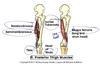
Where do the quadracepts and sartorius originate and insert?

A

Vastus medialis/intermedius/lateralis: originate on femur, inserts in quadracepts tendon (tibial tuberosity)

Rectus femoris: originates below ASIS on hip bone, inserts in quadracepts tendon

Sartorius: orginates at ASIS, inserts in pes anserine (S-G-T)

13
Q

What is the main structure coursing adjacent to the radial sulcus?

A

The radial nerve

14
Q

Which muscles flex the elbow?

A

biceps brachii, brachialis

15
Q

Which muscles extend the elbow?

A

triceps

16
Q

Which muscles supinate the forearm?

A

biceps brachii

supinator

17
Q

Which muscles pronate the forearm?

A

Pronator teres

Pronator quadratus

18
Q

Where do biceps and brachialis insert?

A

Biceps- ulna

Brachilalis- radius

19
Q

What are the compartments of the arm, their innervation and their blood supply?

A

Anterior Compartment (flexor of elbow):

  • Muscles
    • Biceps brachii
    • brachialis
    • coracobrachialis
  • Nerve
    • musculocutaneous
  • Blood
    • brachial artery

Posterior compartment (extensor of elbow)

  • Muscles
    • triceps
  • Nerve
    • radial nerve
  • Blood
    • radial artery
20
Q

What are the contents of the cubital fossa?

A

The median nerve, the brachial artery, the biceps tendon, median cubital vein

21
Q

Sensory distribution of the axillary nerve

A

skin covering deltoid

22
Q

What structures pass through the carpal tunnel?

A

4 tendons of flexor digitorum profunda

4 tendors of flexor digitorum superficialis

tendon from flexor pollicus longus

median nerve

23
Q

What are the borders of the anatomical snuffbox and what does it contain?

A

external tendon: extensor pollicis brevis and abductor pollicis longus

Internal tendon: extensor pollicis longus

containsL radial artery

can palpate scaphoid bone

24
Q

What are the carpal bones?

A

Mnemonic: some lovers try positions// that they cannot handle

Scaphoid, Lunate, Triquetrum, Pisiform

Trapezium, Trapezoid, Capitate, Hamate

(trapezium rhymes with thumb, so its next to the thumb)

25
What are the intrinsic muscles of the hand and how are they grouped?
Thenar: * abductor pollicis brevis * flexor pollicis brevis * opponens Hypothenar: * abductor digiti minimi * flexor digiti minimi brevis * opponens digiti minimi Central muscles * lumbricals * interossei * adductor pollicis
26
What leg compartment is most likely to develop compartment syndrome? What is the treatment?
The anterior compartment. Fasciotomy is the treatment for acute compartment syndrome.
27
Contents passing beneath the flexor retinaculum of the foot? What is this space called?
Tarsal tunnel Tom Dick And Nervous Harry (anterior--\> posterior) * **t**ibialis posterior tendon * tendon of flexor **di**gitorum longus * posterior tibial **a**rtery * tibial **nerv**e * flexor **ha**llucis longus tendon
28
What would you expect with loss of the common fibular nerve?
Supplies lateral (superficial) and anterior (deep) leg compartments, so you would lose eversion (lateral) and dorsiflexion (anterior)
29
Sensory nerve distribution to the sole of the foot (lateral and medial plantar nerves)
Comparable to median and ulnar sensory in the hand: medial supplies medial 3.5 digits, lateral supplies lateral 1.5 digits
30
What is the pectoral girdle?
Clavicle and scapula- its the part of that attaches the upper limb to the trunk
31
What muscles attach to the supraglenoid tubercl, the infraglenoid tubercle and the coracoid process?
supraglenoid tubercle: long head of biceps infraglenoid tubercle: short head of triceps? coracoid: pec minor, coracobrachialis, short head of biceps
32
Course of the cephalic and basillic veins?
note cephalis runs between deltoid and pec major
33
What are the borders of axilla?
34
What are the different parts of the brachial plexus and what spinal roots does it involve
C5-T1 Randy Travis Drinks COld Beer (Roots Trunks Divisions Cords Branches)
35
What spinal levels are these nerves: * musculocutaneous * Median * Ulnar * Axillary * Radial
musculocutaneous: C5,6,7 Median: C5 to T1 Ulnar: (C7,C8,T1) Axillary (C5,6) Radial (C5 to T1)
36
What are the myotomes for: C5-T1
C5: Deltoid (axillary) C6: wrist extension (radial) C7: wrist flexion (median) C8: Finger flexion (median) T1: finger abduction (ulnar)
37
What are the spinal levels for biceps, triceps and brachioradialis reflex?
Biceps: C5 Brachioradialis: C6 Triceps: C7
38
What are the three joints connecting the upper arm to the trunk? What are the 3 ligaments of the A/C joint?
1. Sternoclavicular 2. Acromio-clavicular 1. coracoclavicular 2. acromioclavicular 3. coracoacromial 3. Coraco-acromial
39
In what direction do most shoulder dislocations occur? What ligaments prevent disolocations in other directions?
Inferior Anterior capsular ligaments: superior, middle and inferior glenohumeral ligaments Biceps tendon passes underneath transverse humeral ligament
40
Bursa in the shoulder (x2)
subacromial, subdeltoid....inflammation causes shoulder impingement symptoms
41
What is the arrangement of the rotator cuff muscle tendons around the shouder joint?
Supraspinatus, infraspinatus and teres minor insert on the greater tubercle, subscapularis inserts on the lesser tubercle
42
Ligaments of the elbow
43
Where does the deltoid insert?
On deltoid tuberosity of the humerus
44
What goes through the quadrangular space and the triangluar interval?
45
What are the borders of the trangluar space and the quadragular interval?
46
What are the contents of the cubital fossa? Where do the radial and ulnar nerves pass in relation to this?
47
What is the general arterial supply of the arm?
48
What are the branches of the radial nerve? What are the branches of the musculocutaneous nerve?
radial nerve--\>superficial and deep branches (posterior interosseus) musculocutaneous--\>lateral cutaneous nerve of the forearm
49
What is Lister's tubercle, where is it and what is it for?
50
What the layers of tha anterior forearm?
Deep * pronator quadartus * flexor pollicus longus * flexor digitorum profundus Intermediate: * flexor digitorum superficialis Superficial (originate in common flexor tendon on medial epicondyle) * pronator teres * flexor carpi radialis * palmaris longus * flexor carpi ulnaris
51
Where does brachioradialis insert?
Radius
52
Where does a synovial cyst come from?
enlargement of the synovial sheath surrounding tendons
53
Do Kurt's drawing for the posterior forearm
54
Where is aconeus?
55
What is this showing? What nerve is damaged?
an ape-hand deformity- from damage to the median nerve..would see thenar eminence wasting
56
What are the borders of the anatomical snuff box? What clinically important things are in the snuff box?
Mnemonics: snuff is Excellent, but people who use it should be Abducted (EPL, EPB, APL) Radial nerve, radial artery scaphoid
57
What do the following nerves do in terms of cutaneous and muscle innervation in the arm and forearm: * musculocutaneous * axillary * radial * ulnar * median
Musculocutaneous (C5,6,7) * motor: all the anterior arm muscles * sensory: lateral cuteneous nerve of the forearm Axillary (C5,C6) * motor: deltoid and teres minor (maybe?) * sensory: skin overlying deltoid Radial (C5-T1) * Motor: all muscles in the posterio arm and forearm * Sensory: skin on posterior arm, skin on back of hand lateral 3 digits except fingertips Ulnar (C7,8,T1) * Motor: all intrinsic muscles of the hand * Sensory: medial 1.5 digits (digits 4 and 5) palmar and dorsal skin over these digits via superficial branch Median (C5-T1) * Motor: anterior forearm except 1/2 FDP and FCU * Sensory: lateral 3.5 digits, fingertips dorsally and palmar skin over digits
58
What forms the deep and superficial palmar arches? What parts of the hand are mainly supplied by one of the other?
Ulnar divides into superficial and deep both, radial helps with the deep
59
Thumb actions: extension, flexion, abduction, adduction, opposition
60
What is this showing? Which nerve is injured?
The deep branch of the ulnar nerve (or anything upstream)...if they still have normal ulnar cutaneous innervation, then it is just the deep branch
61
62
What is the course of the ulnar nerve and what are the division in the hand?
superficial (does skin and palmaris brevis) deep (motor to everything...but thenar muscles and 1st and 2nd lumbricals
63
Features of the distal end of the femur
64
ACL and PCL: where they start and where they end
65
Capsular ligaments of the hip joint
66
What artery supplies the head of the femur? The neck of the femur?
Head: obturator via branches through the round ligament of the femur head Neck: medial and lateral circumflex femoral arteries
67
Important bursae in the knee
deep and superficial infrapatellar, prepatellar, pes anserine (deep to tendon)
68
What muscle to the perforating branches of the deep femoral artery pass through?
adductor magnus
69
Where do the posterior thigh muscles orignate and insert
70
Contents of the popliteal fossa
politeal artery and genicular branches tibial nerve common fibular nerva politeal fat and lymph nodes
71
What spinal levels do: hip abduction and adduction?
72
What spinal levels do: hip extension and hip flexion?
73
What spinal levels to: inversion and eversion?
74
What spinal levels do: knee extension and flexion?
75
What spinal levels do dorsiflexion and plantarflexion?
76
What nerves do the sensory of the thigh and leg
lateral cutaneous nerve of thigh (L2,3) Saphenous (medial side of leg and foot) Sural (L5,S1,S2): posterolateral leg and lateral foot
77
Course of great and small saphenous veins
78
Where do hip adductors originate and insert?
All originate from pubis. Pectineus, adductor longus, adductor brevis, adductor magnus all insert on the femur on the linea aspera Gracilis inserts in the pes anserine tendon
79
femoral triangle contents
80
What are the ligaments of the ankle (mortise) joint?
Medial: deltoid ligament Lateral: anterio talofibular ligament, posterior talo-fibular ligament, calcaneal-fibular ligament
81
What are the tarsal bones?
82
What are the joints of the foot?
83
What are the muscles, the function, the innervation and the blood supply of the lateral compartment of the leg?
* muscles * fibularis longus * fibularis brevis * function * eversion * blood supply * peroneal artery * innervation * superficial peroneal nerve (L4-S2)
84
What are the muscles, the function, the innervation and the blood supply of the anterior compartment of the leg?
muscles (all generally dorsiflex the foot) * tibialis anterior (inversion) * extensor hallucis longus * Extensor digitorum longus * fibularis tertiate (eversion) Function: dorsiflexion Blood: anterior tibial artery Nerve: deep peroneal (L4-S2)
85
What are the muscles, the functoin, the innervation and the blood supply of the posterior leg?
Muscles (superficial) * gastrocnemius * soleus * plantaris Muscles (deep) * flexor hallucis longus (toe 1) * flexor digitorum (toes 2-5) * popliteus(unlocks knee) * tibialis posterior (inversion) Function: plantar flexion, flexion of toes Innervation: tibial nerve Blood supply: posterior tibial artery
86
Where do the muscles of the posterior compartment of the leg insert?
The calcaneal tuberosity
87
Course of the sural nerve
88
What is the motor and sensory innervation of the sole of the foot?
\*\*analogous to median and ulnar sensory distribution in the hand
89
What is the blood supply of the sole of the foot?
Posterior tibialis artery passes through the tarsal tunnel and divides into medial and lateral plantar arteries. The lateral plantar arteey forms most of the plantar arch
90
Muscles of the dorsum of the foot, their action, their innervation and blood supply
Muscles: * extensor digitorum brevis (toes 2,3,4) * extensor hallucis brevis (toe 1) Innervation: * deep fibular (just like the anterior compartment!) Blood * dorsalis pedis artery (continuation of anterio tibial a. from anterior leg)
91
What is the femoral artery, what are its major branches
Femoral, deep femoral + perforating branches, goes througha adductor hiatus becomes popliteal, gives off genicular branches, divides into fibular, posterior and anterior tibial, becomes dorsalis pedis (anterior) and lateral and medial plantar arteries (posterior)
92
What is the sensory distribution of the foot?
\*\*in anterior compartment syndrome the deep peroneal nerve will be compressed so the pt should feel everything but the 1st web space