Anatomia Flashcards
La base más o menos circular de las mamas femeninas se extiende transversalmente… y verticalmente…
Transversalmente: desde el borde lateral del esternón hacia línea axilar anterior. Verticalmente: desde la 2.a a la 6.a costillas.
Dos tercios de la mama descansan sobre… que cubre el … ; el otro tercio descansa sobre la fascia que cubre el …
Dos tercios de la mama descansan sobre LA FASCIA PECTROAL que cubre el PECTROAL MAYOR; el otro tercio descansa sobre la fascia que cubre el MUSCULO SERRATO ANTERIOR.
PROCESO AXILAR O COLA (COLA O PROCESO DE SPENCE.)
UNA PEQUEÑA PARTE DE LA MAMA PUEDE EXTENDERSE A LO LARGO DEL BORDE INFEROLATERAL DEL MUSCULO PECTROAL MAYOR HACIA LA FOSA AXILAR Y FORMA EL PROCESO DE SPENCE
LAS GLANDULAS MAMARIAS ESTAN FIRMEMENTE UNIDAS A LA DERMIS DE LA PIEL QUE LAS CUBRE MEDIANTE LOS…
LIGAMENTOS SUSPENSORIOS O DE COOPER ESTOS LIGAMENTOS, ESPECIALMENTE DESARROLLADOS EN LA PARTE SUPERIOR DE LA GLANDULA, AYUDAN A SOSTENER LOS LOBULOS DE LA GLANDULA MAMARIA.
PEZÓN
Se halla en la prominencia más elevada de la mama Rodeado por un área circular pigmentada (la areola)
PARENQUIMA DE LA GLANDULA MAMARIA
ENTRE 15 Y 20 LOBULOS DE TEJIDO GLANDULAR, QUE CONSTITUYE EL PARENQUIMA DE LA GLANDULA MAMARIA
Cada lobulo está drenado por un…, que desemboca independientemente en el peso.
Cada lobulo esta drenado por un CONDUCTO LACTIFERO, que desemboca independientemente en el pezón. Justo por debajo de la areola, cada conducto posee una porción dilatada, EL SENO LACTIFERO
Atlas de Glándula mamaria

Atlas: Arterias de la glandula mamaria

Atlas: Vasos y nodulos (ganglios) linfáticos de la glándula mamaria

Atlas: Drenaje linfático de la mama

Vascularización de la mama
Ramas mamarias mediales de las ramas perforantes
Drenaje Venoso de la mama
se dirige principalmente a la vena axilar, aunque también drena en la vena torácica interna.
IMAGEN: RM SAGITAL QUE MUESTRA LA ESTRUCTURA INTERNA DE LA MAMA Y LAS RELACIONES POSTERIORES.

Imagenes.
Drenajes linfático y vascularización de la mama.
A) Arterias
B) Venas
C) Drenaje linfático.
Drenaje linfático y vascularización de la mama. A) Arterias. B) Venas. C) Drenaje linfático. Los nódulos linfáticos axilares se indican con un asterisco verde.

El drenaje linfático de la mama
La linfa pasa desde el pezón, la areola y los lóbuos de la glándula hacia el plexo linfático subareolar.
La mayor parte de la linfa (> 75%), en especial de los cuadrantes laterales de las mamas, drena hacia los nódulos linfáticos axilares (pectoral, humeral, subescapular, central y apical.)
La mayor parte de la linfa drena inicalmente hacia los nódulos pectorales anteriores.
La linfa de los cuadrantes mediales drena hacia los nódulos linfáticos paraesternales o hacia la mama opuesta.
La linfa de los cuadrantes inferiores puede pasar profundamente hacia nódulos linfáticos abdominales (nódulos frénicos inferiores).
La linfa de los nódulos axilares drena a los nódulos linfáticos supraclaviculares e infraclaviculares, y de estos al tronco linfático subclavio. La linfa de los nódulos paraesternales entra en los troncos broncomediastínicos, que drenan finalmente en los conductos torácico olinfático derecho.
Imagen (cuadrante de las mamas)

Imagen: Venas proximales de la extremidad superior, nódulos linfáticos axilares y drenaje linfático del miembro superior y de la mama.

imagen: Plexo braquial y vasos subclavios en la región cervical lateral (triángulo posterior del cuello).

IMAGEN POR TAC
Lymphatic System
Parietal Nodes
a) Anterior cervical
b) Supraclavicular
c) Subclavicular
d) Interpectoral (Rotter’s) e) Deep axillary
f) Superficial axillary
g) Paramammary
h) Parasternal/Internal mammary i) Infraphrenic
Parietal Nodes
a) Anterior cervical
b) Supraclavicular
c) Subclavicular
d) Interpectoral (Rotter’s) e) Deep axillary
f) Superficial axillary
g) Paramammary
h) Parasternal/Internal mammary i) Infraphrenic

Imagen por TAC
Lymphatic System
Visceral Nodes
j) Para-aortic
k) Paratracheal
l) Upper para-oesophageal
m) Aorticopulmonary
n) Sub-carinal
o) Hilar lymph
p) Mid para-oesophageal
q) Para-vertebral
r) Retrocrural
Visceral Nodes
j) Para-aortic
k) Paratracheal
l) Upper para-oesophageal
m) Aorticopulmonary
n) Sub-carinal
o) Hilar lymph
p) Mid para-oesophageal
q) Para-vertebral
r) Retrocrural

Imagen por TAC
Lymphatic system
Lower Cervical, supraclavicular and sternal notch – Supraclavicular zone
Upper Paratracheal – Upper zone
3a – Prevascular – Upper zone
3p – Retrotracheal – Upper zone
Lower Paratracheal – Upper zone
Subaortic – AP zone
Para-aortic – AP zone
Subcarinal – Subcarinal zone
Para-oesophageal – Lower zone
Pulmonary ligament – Lower zone
Hilar – Hilar zone
Interlobar – Interlobar zone
Lobar – Peripheral zone
Segmental – Peripheral zone
Subsegmental – Peripheral zone
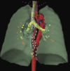
imagen por TAC
Figure 3.5.4 shows the lymph node groups visible at the thoracic outlet. Some of the most commonly irradiated nodes in radiotherapy are the supraclavicular (42), renowned as common routes of spread for head and neck and breast tumours. On the other side of the clavicles can be seen the infraclavicular (43) nodes. The jugular chain (44) lies among the larger blood vessels in this region. The para- oesophageal (45) and paratracheal (46) chains of nodes run vertically along their related structures. The most inferior of the anterior cervical nodes (47) lie in the suprasternal notch.
Figure 3.5.4 shows the lymph node groups visible at the thoracic outlet. Some of the most commonly irradiated nodes in radiotherapy are the supraclavicular (42), renowned as common routes of spread for head and neck and breast tumours. On the other side of the clavicles can be seen the infraclavicular (43) nodes. The jugular chain (44) lies among the larger blood vessels in this region. The para- oesophageal (45) and paratracheal (46) chains of nodes run vertically along their related structures. The most inferior of the anterior cervical nodes (47) lie in the suprasternal notch.

Imagen por TAC
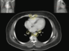
In Figure 3.5.5 (at about the level of T2), the paratracheal and para-oesophageal nodes (50) are closely entwined. In this region, the deep axillary lymph nodes (48) can also be seen. Another common site for lymph node involvement from breast tumours, it can be seen that there is plenty of room for growth in the fatty spaces of the axilla. Anterior and medial to these are the interpectoral nodes (49). These are also known as ‘Rotter’s nodes’. The anterior mediastinal nodes (51) lie just posterior to the sternum.
In Figure 3.5.5 (at about the level of T2), the paratracheal and para-oesophageal nodes (50) are closely entwined. In this region, the deep axillary lymph nodes (48) can also be seen. Another common site for lymph node involvement from breast tumours, it can be seen that there is plenty of room for growth in the fatty spaces of the axilla. Anterior and medial to these are the interpectoral nodes (49). These are also known as ‘Rotter’s nodes’. The anterior mediastinal nodes (51) lie just posterior to the sternum.