Alimentary System Flashcards

1
Q

What is the predominant disease of dogs and cats?

A

Neoplasia

2
Q

What is the predominant disease of horses?

A

Colic

3
Q

Normal oral or gastrointestinal mucosa should be?

A

Smooth and shiny

4
Q

T/F feces can be a window into the health of the alimentary tract?

A

True

5
Q

List four portals of Entry.

A

Ingestion, coughed up and swallowed, hematogenous, migration through the body.

6
Q

What is the most common portal of entry?

A

Ingestion

7
Q

What pathogen is commonly coughed up and swallowed?

A

Rhodococcus equi

8
Q

What is a common parasite that migrates through the body?

A

Spirocerca lupi

9
Q

What defense mechanisms does the alimentary tract have?

A

Saliva, flora, gastric pH, Igs, vomiting, enzymes, phagocytes, high epithelial turnover, increased peristalsis (diarrhea)

10
Q

What species commonly gets cleft palate?

A

Bovine (calves)

11
Q

What are the causes of cleft palate?

A

Genetics, toxins, teratogenic plants (lupines, poison hemlock), materinal exposure to drugs (mares and queens: griseofulvin, primates: steroids)

12
Q

What is cleft palate?

A

Defect in the midline fusion of the palatine shelves.

13
Q

What is the most common complication of cleft palate?

A

Aspiration pneumonia

14
Q

What is a common complication with malocclusions?

A

Difficulties with prehension and mastication

15
Q

What is this called?

A

Cheiloschisis or harelip

16
Q

What is cheiloschisis?

A

Harelip

17
Q

What is brachygnathia?

A

Shorter lower jaw (overbite).

18
Q

What is prognathia?

A

Protrustion of the lower jaw (underbite).

19
Q

Name the layers of the crown of the tooth.

A

Enamel, dentin, pulp.

20
Q

Name the layers of the root of the tooth.

A

Cementum, dentin, pulp.

21
Q

What is dental atrition?

A

Loss of tooth structure due to mastication/chewing.

22
Q

“Step mouth is most common in…?

A

Herbivores

23
Q

What is dental plague?

A

Bacterial film

24
Q

What is dental calculus?

A

Mineralized dental plaque.

25
Q

T/F: Valvular endocarditis can be caused by periodontal disease?

A

True, bacteria follow hematogenous spread and can end up in the heart.

26
Q

Primary diseases of the tongue are rare except for…?

A

Actinobacillosis (Actinobacillus lignieresii)

27
Q

What causes “wooden tongue”?

A

Actinobacillus lignieresii

28
Q

What do you often see clinically in “wooden tongue”?

A

Pyogranulomatous glossitis and sometimes involvement of the cheeks and other soft tissues of the mouth.

29
Q

Gross signs of “wooden tongue” includes..?

A

Tongue sticking out, nodules (represent the pygranulomatous inflammation).

30
Q

What is the etiologic agent of thrush?

A

Candida spp. (especially C. albicans)

31
Q

What is a common cause of thrush?

A

Treatment with antibiotics for long periods of time.

32
Q

What is a characteristic appearance of thrush?

A

A yellowish membrane and yeast and hyphae under the microscope.

33
Q

What causes lingual lesions?

A

Systemic diseases (eg. renal disease, BVD, FMD)

34
Q

What is shown here?

A

Ulcerative and necrotizing glossitis (due to uremia).

35
Q

List 5 causes of stomatitis.

A

Infectious agents, trauma, chemical injury, auto-immune, idiopathic.

36
Q

What type of stomatitis are cats most likely to develop?

A

Lymphoplasmacytic gingivitis.

37
Q

What do cats that have lymphoplasmacytic gingivitis often also have?

A

FeLV or FIV

38
Q

What does feline chronic gingico-stomatitis (FCGS) look like?

A

Oral pain, dysphagia, ptyalism (excessive salivation) and weight loss.

39
Q

What causes feline cronic gingivo-stomatitis?

A

Etiology is unclear, dental plaque, FCV and immune-mediated appear to be involved. It’s also commo in FIV cats.

40
Q

Who most commonly gets chronic ulcerative (lympho-plasmacytic) paradental stomatitis?

A

Older dogs.

41
Q

What is chronic ulcerative paradental stomatitis?

A

Gingival inflammatino (related to plaque) and ulceration of oral mucosa.

42
Q

What is a vesicle?

A

Raised lesion up to 1cm, filled with serous fluid.

43
Q

What is a bulla?

A

A raised lesion larger than 1 cm, filled with serous fluid.

44
Q

What can be ruled out by seeing vesicles in the oral cavity of dogs and cats?

A

Immune-mediated diseases. In cats, often result of calicivirus.

45
Q

What can be ruled out with vesicles in the oral cavity of large animals?

A

Major viral diseases that cause economic loss.

46
Q

What is the pathogenesis of vesicular stomatitides?

A

Viral induced epithelial damage > intracellular edema (balooning degeneration) > vesicles > bullae > rupture > erosion and ulceration.

47
Q

What is this?

A

Vasicular glossitis.

48
Q

What does FMD (Picronavirus) affect?

A

Cloven-hoofed animals (ruminants and pigs). NO HORSES

49
Q

What does vesicular stomatitis (VS, Rhabdovirus) affect?

A

Ruminants, pigs and horses.

Note: Not as bad as FMD, but still have to notify authorities.

50
Q

What does vesicular exanthema of swin (VES, Calicivirus) affect?

A

Only pigs.

51
Q

What does swine vesicular disease (SVD, Enterovirus) affect?

A

Only pigs.

52
Q

Is FMD an exotic disease?

A

Yes.

53
Q

T/F: FMD is highly contagious with high morbidity and high mortality.

A

False, FMD is highly contagious with high morbidity and LOW mortality.

54
Q

How is FMD acquired?

A

Ingesting/inhalation.

55
Q

Where do FMD lesions develop?

A

In areas subjected to mchanical injury.

56
Q

What are the clinical signs of FMD?

A

Excessive drooling (ptyalism) and lameness.

57
Q

What are these pictures of?

A

FMD

58
Q

What is a common finding (and NAVLE question) in young animals with FMD?

A

“Tiger heart”/myocardial necrosis

59
Q

What are the two erosive-ulcerative stomatitides?

A

BVD in cattle and malignant catarrhal fever.

60
Q

What are papular stomatities?

A

Proliferative lesions with coin-shaped papules and ulcers.

61
Q

What causes papular stomatitis?

A

Parapoxvirus (closely related to pseudocowpox).

62
Q

What does contagious ecthyma (orf) cause?

A

Loss of conditino becasue animals neither suckle nor graze (sore mouth). High morbidity, low mortality.

63
Q

What is contagious ecthyma most common in?

A

Lambs and goats 3-6 months of age.

64
Q

Where do you see contagious ecthyma lesions?

A

Lesions develop in sites of trauma.

65
Q

T/F: Contagious ecthyma is not zoonotic?

A

False.

66
Q

What causes oral necrobacillosis?

A

Fusobacterium necrophorum.

67
Q

What clinical signs do you see with necrotizing stomatitis?

A

Characteristic grey-ish caseous necrosis. Nothing else causes this much necrosis.

68
Q

What is this?

A

Necrotizing stomatitis.

69
Q

What is another name for oral necrobacillosis in the calf?

A

Calf diptheria.

70
Q

What is a common sequelae of oral necrobacillosis?

A

Animals can aspirate necrotic tissue.

71
Q

What is a feline eosinophilic granuloma?

A

An ulcerative and granulomatous lesion anywhere in the oral cavity.

72
Q

What does feline eosinophilic granuloma complex include?

A

Eosinophilic granulomas, labial and rodent ulcers.

73
Q

What is this?

A

Eosinophilic granuloma.

74
Q

What causes eosinophilic ulcers?

A

Most likely immune mediated exposure.

75
Q

What is this?

A

Eosinophilic ulcer.

76
Q

What is an eosinophilic granuloma?

A

A proliferative lesion, with granulomatous inflammation with a high number of eosinophils.

77
Q

T/F: Eosinophilic granulomas respond to glucocorticoid therapy.

A

True.

78
Q

Who is most likely to get gingival hyperplasia?

A

Mostly dogs, especially brachycephailc breeds. Major in boxers.

79
Q

What is an epulis?

A

Tumors of periodontal ligament-type stroma.

NOTE: Still controversial if true neoplasm or not.

80
Q

What are fibromatous and ossifying epulis?

A

Benign tumors. The two look similar, but ossifying may have osseous metaplasia.

81
Q

What is an acanthomatous ameloblastoma?

A

Locally agressive apulis, can often recur.

82
Q

Describe canine oral papillomatosis?

A

A transmissible papovavirus-induced papilliform or caulliflower-type lesions. They regress spontaneously and immunity is long lasting.

83
Q

Who is most often affected by canine oral papillomatosis?

A

Animals younger than 1 year-old.

84
Q

Describe an oral papilloma.

A

Thick keratinized stratified squamous epithelium with a pedunculate connective tissue core.

85
Q

In what species is an oral melanoma most common?

A

Dogs

86
Q

Approximately what percentage of oral melanomas in dogs are malignant?

A

90%

87
Q

What are predisposing factors to oral melanoma?

A

Smaller breeds and oral pigmentation.

88
Q

What is the most common location for an oral squamous cell carcinoma?

A

The tongue, can also arise from tonsils.

89
Q

What route would a squamouscell carcinoma metastasize?

A

Lymph.

Remember: Sarcomas tend to go hematogenous first, carcinomas go lymph.

90
Q

What causese congenital megaesophagus?

A

Persistent right aortic arch

91
Q

What breed is predisposed to persistent right arotic arch?

A

German shepherds

92
Q

What age group will you see persistent right aortic arch in?

A

Young dogs (remember, PRAA causes CONGENITAL megaesophagus).

93
Q

What clinical signs will you see with congenital megaesophagus?

A

Weight loss, regurgitation and retching.

94
Q

What is a common cause of death for animals with congenital megaesophagus?

A

Aspiration pneumonia.

95
Q

What is this?

A

Congenital megaesophaguse due to persisten right aortic arch.

96
Q

What can acquired megaesophagus be a manifestation of?

A

Myasthenia gravis.

97
Q

What is a common cause of death with acquired megaesophagus?

A

Aspiration pneumonia.

98
Q

What is choke?

A

Foreign material completely obstructs the lumen of the esophagus, compression necrosis can be seen.

99
Q

What are two common locations for choke to occur?

A

Dorsal to the larynx and in the thoracic inlet.

100
Q

What will you see with an animal that has choke?

A

Can’t eat, drink or eructate (bloat!!), see retching, hypersalivation (ptyalism), and regurgitation (if the blockage is in the thoracic inlet and ingesta can get that far)

101
Q

What do you see on necropsy in cases with acid reflux?

A

Linear ulceration of the esophageal mucous membrane.

102
Q

What is a common cause of ulcerative erosions in the esophagus?

A

BVD

103
Q

What do you see on necropsy with ulcerative erosion of the esophagus?

A

Small, oval-shaped ulcers.

104
Q

What is parasitic esophagitis?

A

Chronic, granulomatous esophagitis caused by Spirocerca lupi.

105
Q

What else can you see with parasitic esophagitis?

A

Tumour development (osteous and fibrous).

106
Q

What clinical signs will you see with parasitic esophagitis?

A

Dysphagia, painful swallowing, etc.

107
Q

What is rumenal tympany?

A

Bloat

108
Q

What is the cause of primary rumenal tympany?

A

New diets (usually high in CHO) that promote formation of stable foam.

109
Q

What causes secondary rumenal tympany?

A

Physical or functinoal obstruction of the esophagus resulting in failure to eructate.

110
Q

How does bloat cause fatality?

A

Rumen compresses the diaphragm causing cardiovascular collapse and death.

111
Q

How can you tell in necropsy if the bloat was pre- or post-mortem?

A

Check rumenal contents for bubbles, and check rumen pH (too much CHO will decrease the pH).

112
Q

What is the most reliable post-morten indicator of ante-mortem bloat?

A

Bloat line in the esophagus at the thoracic inlet.

113
Q

What is this?

A

A bloat line

114
Q

What is a common name for traumatic reticulitis?

A

“Hardware disease”

115
Q

What is common to see as a complication of hardware disease?

A

Chronic pericarditis.

116
Q

What causes chemical rumenitis?

A

Feed with high grain or CHO which causes lactic acidosis.

117
Q

What are “stellate ulcers”?

A

Rumenal scars, likely from a bout of chemical rumenitis that has healed.

118
Q

What is a common sequel to chemical rumenitis?

A

Mycotic rumenitis.

119
Q

What species do you see GDV in?

A

Dogs and swine?

120
Q

What organ is also often involved in a GDV?

A

The spleen

121
Q

When are you most likely to see GDV in swine?

A

After excessive feed and water intake, followed by physical activity in a competitive group situation.

122
Q

What clinical signs will you see with a dog that had a GDV?

A

Abdominal distension, non-rpoductive retching, hypersalivation, restlessness.

123
Q

What is this?

A

GDV

124
Q

What cattle are most susceptible to DA?

A

Post-parturient dairy cows and calves

125
Q

What side is the most common displacement for an abomasum?

A

Left side.

126
Q

T/F: LDA is fatal.

A

False, LDA is generally non-fatal.

127
Q

What do 20% of RDA result in?

A

Abomasal volvulus.

128
Q

What clinical signs will you see with a displaced abomasum?

A

adbominal pain, elevated heart rate, anorexia, dehydration, depressed peristalsis with lack of feces and abomasal tympany.

129
Q

What can be done to prevent a displaced abomasum in prone animals?

A

Gastropexy

130
Q

What species is most likely to have gastric impaction/rupture?

A

Horses

131
Q

What causes gastric impaction/rupture?

A

Intestinal obstructions (ileus) leading to an adynamic (paralytic) or mechanical ileus.

132
Q

How can you tell if a gastric rupture is post-mortem?

A

You won’t see evidence of hemorrhage if it’s post-mortem.

133
Q

What is seen in pigs that are fed finely ground rations?

A

Gastric ulceration.

134
Q

What is this?

A

Gastric ulcers.

135
Q

What can cause gastric ulcers in a horse?

A

NSAIDs, because they decrease prostaglandin production which is important for the perfusion and protection of the gastric mucosa.

136
Q

T/F: Most gastric ulcers in dogs and cats are idiopathic?

A

True.

137
Q

What lesion in small animals can lead to gastric ulcers?

A

Mast Cell tumors. Histamine binds the receptors on parietal cells increasing HCL.

138
Q

What stain is used for mast cells?

A

Toluene blue.

139
Q

What are two major causes of gastric inflammation?

A

E. coli and Salmonella.

140
Q

What is another major cause of gastric inflammation?

A

Urea (uremic gastritis, will feel gritty upon sectioning).

141
Q

What is characteristic of uremic gastritis, and renal disease?

A

White-ish discolouration and mineralization of the lamina propria.

142
Q

What is a common name for abomasal gastritis?

A

Braxy or Bradsot

143
Q

What causes Braxy?

A

C. septicum

144
Q

When does Braxy commonly happen?

A

In the winter, it’s associated with feeding frozen feed.

145
Q

How does the abomasum appear with Braxy?

A

Red, swollen tissue with mucus and some fibrin. Also will notice gasses in the tissue due to the Clostriddium (feels like crepitus).

146
Q

What is this?

A

Abomasitis, Bacterial Gastritis, Braxy or Bradsot

147
Q

What is a cause of mycotic abomasitus?

A

A sequel of long-term Ab use.

Causes death of resident flora and allows angio-invasive fungi to grow.

148
Q

What do you commonly see causing mycotic abomasitis?

A

Aspergillus, Absidia, Rhizopus, Mucor spp.

149
Q

What is this?

A

Mycotic abomasitis.

150
Q

What can result from mycotic abomasitis?

A

Mycotic vasculitis/perivasculitis. Fungi invade the tissues and enther the blood vessels. Can result in vasculitis and thrombosis.

151
Q

What is this?

A

Mycotic vasculitis.

152
Q

What is a common cause of parasitic gastritis?

A

Gasteropilus nasalis and intestinalis.

153
Q

What do bots do to the lining of the stomach?

A

Create erosion and ulceration.

154
Q

What is this?

A

Erosive-ulcerative lesions caused by G. intestinalis.

155
Q

What will you see with gastritis due to Ostertagia spp.?

A

Proliferative abomasitis with a moroccan leather appearance of affected mucosa.

156
Q

What do you see in cases of Draschia megastoma infestations in horses?

A

Brood poud cloes to margo plicatus and a granulomatous gastritis.

157
Q

What is a common sign of Haemonchus contortus in sheep?

A

Bottle jaw, anemia nad hypoproteinemia due to the parasitic abomasitis.

158
Q

What is a common gastric neoplasia of horses?

A

Gastric carcinomas.

159
Q

Where do gastric carcinomas in horses usually occur?

A

In the esophageal portion of the stomach.

160
Q

What else can a gastric carcinoma in a horse cause?

A

Can cause peritoneal carcinomatosis.

161
Q

What is this?

A

Peritoneal carcinomatosis.

162
Q

What do you see in cases of gastric lymphoma in a horse?

A

Nodules in the stomach, not confined to the margo plicatus (tells you that it’s not megastoma, plus no parasites present).

163
Q

What will you see in a case of lymphosarcoma in a cow?

A

Thickened, nodular abomasal folds, and also ulceration. Cows may also have diarrhea with some melena present.

164
Q

What is the most common segmental anomaly of the intesting of domestic animals?

A

Atresia coli

165
Q

What is atresia coli?

A

Complete occlusion/obliteration of the intestinal luman.

166
Q

What is atresia ani?

A

Lack of a perforation in the anus.

167
Q

What is lethal white syndrome in foals?

A

Congenital colonic agangliosis.

168
Q

What do you most commonly see lethal white syndrome in?

A

Completely white foals with blue eyes.

169
Q

What breed is lethal white syndrome in foals most associated with?

A

American paint horses.

170
Q

What is lethal white syndrome?

A

It’s a lack of parasympathetic ganglie in the colon and cecum giving GI immotility. Foals start to develop colic because of the meconium in the cecum.

171
Q

What do you see pathologically with lethal white foal syndrome?

A

Absence of myenteric and submucosal parasympathetic ganglia in the wall of the ileum, cecum and colon leading to intestinal immotility and colic.

172
Q

What is an enterolith and what does it cause?

A

Mineral deposits around a nucleus that can cause a complete gastric obstruction.

173
Q

What are the most common minerals that make up an enterolith?

A

Magnesium, ammonium phosphate (struvite).

174
Q

What is the nucleus for enterolith formation?

A

A foreign body.

175
Q

What is a trichobezoar?

A

A hair ball.

176
Q

What large animal species most commonly get trichobezoars?

A

Cattle.

177
Q

Where are trichobezoars usually found?

A

Forestomach and abomasum.

178
Q

What is a phytobezoar/phytotrichobezoar?

A

Composed mostly of plant material impregnated with some phosphate salts. Can be found in horses.

179
Q

What is this?

A

Ascarid impaction.

180
Q

What is an acquired stenosis?

A

It’s a stricture as a result of a penetrating wound. Lumen heals with scar tissue which decreases the lumen diameter.

181
Q

What is rectal stricture in pigs most commonly associated with?

A

Salmonellosis.

182
Q

What is a hernia?

A

Protrusion of an organ or part of an organ/tissue through an abnormal opening.

183
Q

What are the 2 types of hernia?

A

Internal and external.

184
Q

What is an internal hernia?

A

Displacement of intestine through a normal or abnormal foramina within the abdominal cavity (rare).

185
Q

What is an external hernia?

A

Displacement of loops of intestinge, omentum and occasionally other viscera (hernial contents) outside the abdomenal cavity.

186
Q

What is an eventration?

A

When the dispalced abdominal contents are not covered by parietal peritoneum or skin.

187
Q

What is this?

A

An eventration.

188
Q

What are two causes for the formation of a diaphragmatic hernia?

A

Trauma (eg. HBC) congenital.

189
Q

What is this?

A

Scrotal hernia.

190
Q

What can incarceration of a piece of small intesting lead to?

A

Venous infarction, sepsis and death.

191
Q

What is this?

A

Torsion of the left colon.

192
Q

What can happen to a colonic torsion during necropsy?

A

The torsion can come undone! Look for a line that indicates congestion.

193
Q

How can you tell that a horse died from a colonic torsion, even if no torsion can be seen on necropsy?

A

Line of congestion.

194
Q

What is this?

A

Line of congestion indicating colonic torsion.

195
Q

What is this?

A

Intestinal volvulus.

196
Q

What happened here?

A

Herniation of small intestine through the epiploic foramen (internal hernia).

197
Q

T/F: A penduculated lipoma can cause intestinal strangulation.

A

True, the peduncle can wrap around the intestines because the peduncle can be very long.

198
Q

What can happen with LARGE pedunculated lipomas?

A

You can see them twist around themselves and become necrotic mineraize.

199
Q

What is an intussusception?

A

One piece of gut telescopes into another piece.

200
Q

What is the most common direction of an intussusception?

A

Normally more cranial will telescope into the more caudal portion.

201
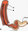
Q

Which is the intussusceptum and which is the intussuscipiens?

A

A: intussuscipiens

B: intussusceptum

202
Q

What is this?

A

Intussusception.

203
Q

What are the 3 major causes for intestinal inflammatory diseases?

A

Viral, bacterial and parasitic.

204
Q

What do you commonly see clinically in intestinal inflammation?

A

Diarrhea causing dehydration, acidosis, malabsorption, hypoproteinemia, and electrolyte imbalances.

205
Q

What is the most diagnostic (and most sever form) of BVD?

A

Peyer’s patch necrosis.

206
Q

What lesions do you see associated with BVD?

A

Erosive ulcerative lesions anywhere in the oral cavity and intestinal tract plus Peyer’s patch necrosis.

207
Q

What is the most common age group to be affected by BVD?

A

6 months to two years of age.

208
Q

What causes malignant catarrhal fever?

A

Gamma herpesvirus.

209
Q

What type of lesions do you see in cases of malignant catarrhal fever?

A

Fibrino-necrotizing vasculitis.

210
Q

What type of malignant catarrhal fever is present in the US?

A

tHe, “Shee-associated MCF”

211
Q

What is a common cause of neonatal diarrhea in calves?

A

Coronavirus infections. Can be combines with Rotavirus or Cryptosporidium.

212
Q

What other viruses can combine with coronavirus to cause neonatal diarrhea in calves?

A

Rotavirus or Cryptosporidium.

213
Q

What sort of gut morphology do you see microscopically with transmissible-gastro-enteritis?

A

Severe villous atrophy and fusion.

214
Q

What does rotavirus cause in young animals?

A

Diarrhea.

215
Q

How does rotavirus cause diarrhea in young animals?

A

Damages the surface enterocytes causing variable degrees of villous atrophy.

216
Q

What species often has subclinical infections of rotavirus?

A

Swine (piglets).

217
Q

What can be seen in parvovirus enteritis?

A

Some cases have Peyer’s patch necrosis like BVD cases.

218
Q

In cats, what causes parvovirus enteritis?

A

Feline panleukopenia.

219
Q

How can you get a firm diagnosis of parvovirus?

A

TEM with a fecal sample.

220
Q

How does FIP often present?

A

In young cats with abdominal distention due to exudate into peritoneal space.

221
Q

What can you see in the peritoneal space of a cat that has the wet form of FIP?

A

Commonly will see fibrin plaques all over the surface of the intestines.

222
Q

What can you see on the kidney of a FIP cat that usually is a good sign that the cat actually has dry FIP?

A

Nodules that follow the blood vessels.

223
Q

What is an important cause of enteritis in neonatal animals?

A

E. coli

224
Q

What is enterotoxemic colibacillosis?

A

Edema disease in pigs.

225
Q

What causes edema disease?

A

A bacterial enterotoxin that causes endothelial cell injury in arterioles resulting in fluid loss and edema.

226
Q

What can you see in edema disease?

A

Focal, bilateral symmetric encephalomalacia and edema in the mesocolon (spiral colon).

227
Q

Which type of Clostridium perfringens is the most common?

A

Type D

228
Q

What animal group does Clostridium affect?

A

The best nourished animals.

229
Q

What do you see in animals with C. perfringens.

A

Animals may be found dead, or may exhibit bloody diarrhea.

230
Q

What does C. perfringens produce?

A

Angiotoxin.

231
Q

What do you see in animals affected by C. perfringens?

A

Intestinal lesions and focal symmetrical encephalomalacia (FSE) in sheep.

232
Q

What can you see microscopically with clostridial enteritis?

A

Necrotic villi lined by G+ bacilli, hypereosinophilia.

233
Q

What diesase does Clostridium piliforme cause and what organs does it effect?

A

Tyzzer’s disease, which targets the liver and causes lesions in the intestines (port of entry) and the heart.

234
Q

What does Clostridium perfringens type A and Clostridium difficile cause?

A

Colitis X/Typhocolitis of horses.

235
Q

What diseases can salmonellosis cause?

A

Enterocolitis to septicemia.

236
Q

T/F: Salmonella is not a cause of zoonotic and nosocomial infection.

A

False, it IS a cause of zoonotic and nosocomial infection.

237
Q

What are common findings with Salmonellosis?

A

Ulcerative and fibrino-necrotizing enterocolitis. Intestinal contents are malodorous and contain mucus, fibrin and occasionally blood. Feces has a “septic takn odor”.

238
Q

What is this?

A

Edema and hemorrhage due to Salmonellosis with fibrino-necrotizing entero-colitis.

239
Q

What is a common sequel of Salmonellosis?

A

Embolic mycotic pneumonia.

240
Q

What is this?

A

Embolic mycotic pneumonia.

241
Q

What is this?

A

Embolic mycotic pneumonia.

242
Q

What is this?

A

Button ulcers in a chronic salmonellosis case.

243
Q

What disease is caused by Lawsonia intracellularis?

A

Porcine Proliferative Enteropathy (PPE)

244
Q

What age group is most commonly associated with Porcine Proliferative Enteropathy?

A

Weaner and grower pigs.

245
Q

What can you see clinically with Lawsonia infections in pigs?

A

Proliferative ileitis, and necrotic enteritis.

246
Q

What is this?

A

Proliferative hemorrhagic enteropathy from L. intracellularis.

247
Q

What stain can you use to confirm a diagnosis of Lawsonia intracellularis?

A

Silver stain.

248
Q

What organism causes swine dysentery (spirochetal colitis)?

A

Brachyspira hyodysenteriae.

249
Q

What age group does Brachyspira affect?

A

Pigs 8-14 weeks of age.

250
Q

How is swine dysentry characterized?

A

Large bowel diarrhea with mucous and blood in the feces.

251
Q

What does Rhodococcus equi cause in young horses?

A

Enterocolitis.

252
Q

What type of lesions are seen with R. equi?

A

Suppurative pyogranulomatous pneumonia in foals and also enteric, ulcerative and pyogranulomatous lesions with prominent regional lymphadenitis.

253
Q

What is this?

A

Pyogranulomatous colitis due to Rhodococcus equi.

254
Q

What is this?

A

Pyogranulomatous colic lymphadenitis.

255
Q

What is this?

A

Rhodococcus equi pneumonia.

256
Q

What is Johne’s disease?

A

Paratuberculosis.

BONUS POINT: Mycobacterium avium ssp. paratuberculosis

257
Q

What is Johne’s disease?

A

A chronic disease of ruminants with diarrhea, emaciation and hypoproteinemia.

258
Q

What age group does Johne’s disease affect?

A

Animals older than 19 months.

259
Q

What is this?

A

Granulomatous enteritis.

260
Q

What is this?

A

Granulomatous lymphangitis with lymphangiectasia.

Note: the thickened lymphatic vessels which shows granulomatous lymphangitis, should be Johne’s.

261
Q

What dog breed can you commonly see granulomatous colitis in?

A

Boxers

262
Q

T/F: You can commonly see lesions of granulomatous colitis on the serosal surface.

A

True.

263
Q

What causes granulomatous colitis in Boxers?

A

Selective intramucosal colonization by specific strains of E. coli.

264
Q

How can cryptosporisiosis be diagnosed?

A

By finding numerous protozoal organisms attached to the apical surface of enterocytes.

265
Q

What is this?

A

Coccidial enteritis.

266
Q

What is this?

A

Eimeria spp.

267
Q

What blood sucking parasites cause anemia and hypoproteinemia?

A

Hookworm

268
Q

What parasite causes “milk spotted livers”?

A

Ascaris suum.

269
Q

What species are intestinal tumours most common in?

A

Dogs and cats.

270
Q

What are most primary tumors?

A

Carcinomas.

271
Q

Where do most lymphosarcomas arise from?

A

The intestines.

272
Q

What are most cases of lymphosarcomas manifestations of?

A

Multicentric lymphosarcomas.

273
Q

What is the most common neoplasm in cats?

A

Lymmphosarcoma, ailimentary form especially.