Adult Chest Flashcards
Mnemonic for DDx of a lung cavitation
CAVITY
- Cancer: usually squamous
- Autoimmune: Wegeners, rheumatoid/Caplan syndrome
- Vascular: septic/bland emboli (Lemierre)
- Infection: TB (primary progressive or reactivation), mycobacterial
- Trauma: pneumatoceles
- Young/congenital: CPAMs, sequestrations
Which structure is responsible for a juxtaphrenic peak?

The inferior pulmonary ligament.
- The juxtaphrenic peak is assoc w/upper lung volume loss of any cause.
What forms the medial border of the R paratracheal stripe?

R tracheal wall.
- The medial pleura forms its lateral border.
- If it’s widened (>4mm) then it’s often due to lymphadenopathy.
- Other causes: mediastinal hemorrhage, mediastinal mass, vessel enlargement.

What’s the name of the fissure that separates the medial basal segments from the other basilar segments (in both lungs)?
Inferior accessory fissure.
- It’s typically vertical & complete.
- More commonly seen on the R.
- The superior accessory fissure separates the lower lobe superior segment from the basilar segments.

Which LN station is this?
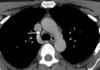
- R lower paratracheal (4R).

Which airway level is the first to lack cartilage in its wall?
Terminal bronchiole.
- Respiratory bronchioles branch off these & so are the 2nd airway to lack cartilage.
- These are the first airways to contain alveoli.
- They terminate as alveolar ducts.

In the intercostal spaces, what is the cranial to caudal order of the neurovascular bundle?
VAN: vein, artery, nerve.

Into what structure does the thoracic duct typically drain?
L subclavian vein + IJV confluence.

What parts of the body does the thoracic duct not drain directly or indirectly?
- R upper extremity & chest.

In which decade of life should there be complete fatty replacement of the thymus?
Eighth! So it’s entirely fatty by age 80.
- Don’t mistake a normal thymus for a thymolipoma, as the latter will have a discrete mass, containing fat (see below).
- A normal thymus will appear strandy.

Name the muscle.

Supraspinatus
- The scapula creates a “T” shape dividing the muscles into subscapularis deep (or anterior) to the scapula, infraspinatus lateral and inferior to the spine of the scapula, and supraspinatus medial and superior to the spine of the scapula. Teres minor courses along the lateral aspect of infraspinatus.
Which connective tissue disease is most highly associated w/obliterative bronchiolitis?
Rheumatoid arthritis.
- Obliterative bronchiolitis = air trapping.

Pulmonary infarction typically involves which vessels?
- Pulmonary arteries.
- You know this b/c of PEs, which travel to the lungs via PAs.
- PEs & pulmonary involvement of vasculitis are the most frequent causes.
DDx for tree-in-bud opacities & why?
DDx: endobronchial spread of infection (TB) & aspiration.
- It represents filling of the branching distal airways, and the centrilobular nodule/airway.
What is the Monod sign?
- Fungal ball within a pre-existing cavity = aspergilloma.
- Frequently the mass is gravity dependent & can move.

Name of this sign in the setting of PE?

Hampton hump
- Peripheral consolidation of a pulmonary infarct.
With what exposure is pulmonary alveolar proteinosis associated?

- Silica.
- It can also be idiopathic.
What is the name of this sign and what disorder is most closely related?

- Galaxy sign
- Sarcoid
What is the max size limit of a pulmonary bleb?
- 1cm.
- Bulla >1cm

At what CD4 count does PJP begin to occur in HIV pts?

<200
- CMV appears in sicker pts, CD4<100.
- So does aspergillus & nontuberculous mycobacterial, as well as cryptococcus, disseminated histoplasmosis.
- Ix findings of PJP & CMV are similar.
MRSA infection:
- What is a risk factor for community-acquired infection?
- What abx are used to treat it?

- Incarceration.
- Ceftazidime & vancomycin: broad-spectrum.
What is the most common infectious agent to cause pulmonary septic emboli?
- S. aureus
- May see the “feeding vessel” sign, which is a vessel supplying the infarcted parenchyma. This can also be seen in cancer.
What is the next step in mgt for an immunocompromised patient with an acute respiratory illness and an abnormal chest radiograph with multiple, diffuse, or confluent opacities?
- CT chest w/o IV contrast.
- According to ACR Appropriateness Criteria.
Dx? Pt who underwent heart xsplant 3 months prior.

Dx: aspergillis, “halo sign”.
- In the setting of heart transplant, new pulmonary parenchymal nodules are more likely infectious than neoplastic, though both should be considered depending on the clinical context. In this case, there is a solid nodule with surrounding ground-glass opacity, known as the halo sign. This sign is associated with invasive fungal infection, especially angioinvasive aspergillosis, which is a common opportunistic infection following transplant. CMV pneumonia much more commonly presents with diffuse ground-glass opacities than an isolated discrete nodule. There are no features to suggest rounded atelectasis in this case.
Posttransplant lymphoproliferative disease (PTLD) should be considered for nodules/masses in a posttransplant patient; however, it typically occurs at least 1 year after transplantation. Nodular disease prior to that time frame is much more likely to be infectious. PTLD is also commonly accompanied by intrathoracic lymphadenopathy.


