9.11 Nerves of the Lower Limb Flashcards
• The origin of the lumbosacral plexus and the formation of its major branches • The origin, course and distribution of the sciatic, femoral, obturator, common fibular and tibial nerves including the muscles and muscle groups that each supplies and their sensory distribution • The common sites of peripheral nerve injury and the possible functional effects of such damage • The dermatomes and myotomes of the lower limb
From what spinal roots does the lumbar plexus derive its supply from?
The lumbar plexus is derived from spinal nerve roots L1-5
What are the 6 main terminal branches of the lumbar plexus?
- Iliohypogastric Nerve
- Inguinal Nerve
- Genitofemoral Nerve
- Lateral Femoral Cutaneous Nerve
- Obturator Nerve
- Femoral nerve
(in order of their branching off the plexus)

Describe the path of the lumbar plexus nerves from their origin to the lower leg
The lumbar spinal nerves emerge from interverebral foramen and pass down to the periphery and pass into psoas major.
They converge on psoas and reform within the substance of psoas and come out either..
- 4 lateral to psoas
- 1 branch medial to psoas (obturator nerve)
- 1 branch goes through the substance of psoas (genitofemoral)

Do the ventral rami or dorsal rami of L1-L4 make up the lumbar plexus?
Only ventral rami of the spinal cord (dorsal rami never form plexuses)
What is special about the L2,3,4 divisions of the lumbar plexus?
They have both an anterior and posterior division (like contributions of the brachial plexus of the arm)
(L1 and L5 only have anterior division)
This anatomy relates to embryolgical devleopment.

Explain what the anterior division of the Lumbar plexus generally innervates.
Relate this to embryolgical development of the lower limb.
In development, the lower limb is buds out externally rotated and it rotates 90 degrees inwards/medially in utero.
Thus before this rotation happens:
- the adductor muscle compartment is originally anterior, thus this region is supplied by the anterior division.
- the posterior division thus supplies what is now laterally (used to be posterior) which is the abductors (femoral)

What does the lumbar plexus innervate?
Lumbar plexus is primarily distributed to the lower limb.
- The termination of thoracic nerve, T12 forms some of the lumbar plexus and also contributes to nerves to the inguinal region
- L1 also supplies the lower abdominal wall
Which nerves come out of the following …
- Lateral Border of the Psoas?
- The Anterior Border of Psoas (through it)?
- Medial Border of Psoas?
Draw them coming out
Lateral
- Iliohypogastric (L1)
- Ilioinguinal (L1)
- Lateral Femoral Cutaneous Nerve (L2,3)
- Femoral Nerve (L2,3,4)
Anterior
- Gentiofemoral (L1,2)
Medial
- Obturator (L2,3,4)

Describe the path of the nerves coming out of L1
The Ileioinguinal nerve and Ileohypogastric nerves go to the lower parts of the anterior abdominal wall (transverse oblique and transverse abdominus) and the pelvic region
What is the relationship between the ileohypogastric nerve and ileoinguinal nerve?
Ileohypogastric is the L1 nerve itself and has the same exit pathway as a typical intercostal nerve.
- It comes out and supplies the skin of the anterior abdominal wall by cutaneous branches
The ileoinguinal nerve it is a colateral branch of the ileohypogastric.
- It itself has no collateral branches emerging from it, thus it doesn’t have cutaneous supply

Describe how the lateral femoral cutaneous nerve gets through the pelvis
- What is special about it?
- Describe how pathology can occur to it
It passes…
- Under the inguinal ligament about a 1cm medial to the ASIS.
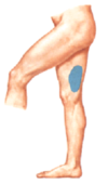
- It is a purely sensory nerve supplying skin over the lateral aspect of the thigh.
- It is susceptible to compression at the site where it passes under the inguinal ligament particularly during pregnancy, overweight/obese as the increased weight can compress the nerve = MARALGIA PARASTHETICA

What makes up the borders of the femoral triangle?
- Laterally Sartorius Muscle
- Anteriorly Inguinal Ligament
- Medially Adductor Longus Muscle

There are vascular structures that pass through the femoral triangle on their way to the lower limbs.
What are these structures?
What are they protected by?
Artery, vein and canal - which holds lymph nodes)
They are surrounded by the deep fascia called the femoral sheath

How and where does the femoral nerve pass through in the femoral triangle (in relation to the femoral sheath)
The femoral nerve passes into femoral triangle below inguinal ligament. It is lateral to the sheath, NOT inside the sheath.

Describe the order of the vessels running through the femoral artery (medially to laterall)
Lateral to Medial:
- Nerve (femoral)
- Artery
- Vein
- (Lymph vessels)
How to remember: “N.A.V.igate” to the good stuff

What happens to the femoral nerve as soon as it enters the femoral triangle?
As soon as it enters the triangle (few cm below) it breaks up into its branches most of which are muscular but also some sensory.
This division is into the superficial and deep branches

Describe the femoral nerve in terms of:
- What spinal nerve roots
- Anterior or posterior division
- Emergence from Psoas Major Muscle
- L2,3,4
- Posterior Division
- Emerges from the lateral border of psoas
What landmark separates the superior branches of the femoral nerve from the deep branches? What does this structure do?
The lateral circumflex femoral artery.
Branches of this artery go up to head of femur to supply the neck.

Describe the path of the femoral nerve descending down the leg
The femoral nerve is the largest branch of the lumbar plexus.
- Descends in the abdomen through the psoas major muscle.
- Travels through the pelvis to approximately the mid point of the inguinal ligament. It then traverses under the inguinal ligament into the thigh and splits into an superficial (anterior) and deep (posterior) division.
- It passes through the femoral triangle lateral to the femoral vessels and gives off articular branches to the hip and knee joints.
- These vessels are deeply embedded in the thigh

Where is the femoral nerve most suseptible to injury?
Once it gets into the thigh it is embedded in muscle, it most susceptible to injury in the groin as it passes through inguinal ligament.
It is not commonly associated with typical injury but damage to it leads to loss of motor control to quadricepts, lost sensory and diminished patellar tendon reflex.
What motor innervation does the femoral nerve supply?
Do these nerves come from the superficial or deep divison?
Femoral supplies motor innervation to the muscles located in the anterior compartment of thigh
- Quadriceps
- Sartorius
- Pectineus
Superficial gives rise to one muscle: sartorius. (Everything else is sensory to the thigh).
Deep branch supplies the remaining muscles of the anterior: quads and pectineus. (And gives off one sensory branch: saphenous nerve) to the medial aspect of leg and foot
What sensory innervation does the femoral nerve give?
Do these nerves come from the superficial or deep division?

It supplies sensory innervation to the medial aspect of the thigh via the saphenous nerve
As it crosses the joint, it also supplies articular branches to the hip and knee joint.
Superficial gives rise to one muscle: sartorius. (Everything else is sensory to the thigh).
Deep branch supplies the remaining muscles of the anterior: quads and pectineus. (And gives off one sensory branch: saphenous nerve) to the medial aspect of leg and foot terminating at the first metatartsal-phalageal joint

Describe the obturator nerve in terms of…
- Lumber Spinal Roots
- Anterior or posterior division of the plexus
- Origin in relation to psoas
- Its passage through the pelvis to the leg
- L2,3,4
- Anterior divisions
- Medial border of Psoas
- Passes down the lateral wall of pelvis → through the obturator canal → medial compartment of thigh
The obturator nerve divides into 2 divisions (Anterior and posterior). What separates them?
What does the nerve supply?
It divides into an anterior and posterior division in relation to ADDUCTOR BREVIS muscle.
- Anterior division supplies motor to the superficial adductor muscles
- Posterior division supplies motor to fibres of the adductor magnus (from both obturator nerve and sciatic nerve)
- Articular branches to the hip