3. General Dermatology Flashcards
Worldwide prevalence of psoriasis?
2%
What percentage of psoriatic patients develop symptoms of psoriatic arthritis(PsA) ?
5-30%
Psoriasis age peak & distribution
Bimodal distribution
Peaks at 20-30 & 50-60 yrs
Name some genetic factors of psoriasis
- PSORS-1 susceptibility locus (on chromosome 6p)
- HLA- Cw6
HLA - B27 is ass/w ?
Sacroilitis- associated psoriasis
PsA
Pustular psoriasis
HLA - Cw6 & Psoriasis
- 10–15 times ↑risk
- Positive in 90% of early-onset psoriasis
- 50% of late onset
- strongly a/w guttate psoriasis
Strongest HLA risk factor for early-onset disease?
HLA-Cw6
( Cw6> B57,DR7 )
HLA ass/w with guttate and erythrodermic psoriasis?
HLA B13 & B17
HLA associated with palmoplantar pustulosis
HLA-B8, Bw35, Cw7, and DR3
Pathogenesis of psoriasis
Primarily T-cell disorder
- CD8+ in epidermis
- mix of CD4+/CD8+ in dermis
- Increased Th1 cytokines, IL-1, IL-6, TNF-a
- Decreased IL-10
- ↑dendritic cells in psoriatic skin
Triggering factors of psoriasis
- External: Trauma (Koebner phenomenon)
- Internal:
- Infections ( streptococcal pharyngitis n.1, HIV)
- Endocrine factors
- Stress
- Drugs
- Obesity
- Smoking, alcohol consumption
Triggering factor in generalized pustular psoriasis?
Hypocalcemia
Triggering factor in impetigo herpetiformis?
Pregnancy
MC drugs that can exacerbate/trigger psoriasis
- Lithium
- IFNs
- β-blockers
- Antimalarials
- TNF-a inhibitors
- CS tapers in pustular psoriasis
Length of latency period btw trauma (Koebner) and appearance psoriatic lesions?
2-6 weeks
TNF-a inhibitors may induce which type of psoriasis?
Plaque psoriasis
+/- palmoplantar pustulosis
Latency period btw drug initiation & psoriatic skin eruption
- Short latency (<4 weeks): terbinafine, NSAIDs
- Intermediate latency (4 to 12 weeks):
antimalarials, ACEIs - Long latency (>12 weeks): β-blockers,
lithium
Dx?

Chronic plaque psoriasis
Sharply demarcated, erythematous, scaly plaques

Dx?

Multiple large plaque psoriasis
Obvious symmetry of the plaques on the upper extremities
+/- pruritus & hemorrhagic crusts due to scratching
Dx?

Annular plaques of psoriasis due to central clearing
Dx?

Sunburn related Koebner phenomenon
Dx?

Guttate psoriasis
Dx?

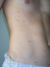
Linear Koebner
+ widely scattered guttate lesions
Dx?

Generalized pustular psoriasis
Broad areas of erythema with numerous pustules & formation of lakes of pus