2nd Week Discussion Flashcards
clinical features of erythema multiforme (3)
Clinical features EM minor: –Skin (extremities) –Mucosa (oral, conjunctival, genitourinary, respiratory) –Hemorrhagic crusting of vermilion zones
ID

Erythema multiforme
describe the erythema multiforme lesions on skin (2)
Variety of appearances “multiforme”
• Round, dusky-red patches on skin of
extremities “target lesions
” • Bullae with necrotic centers

Erythema multiforme (EM)
• Erythematous patches oral mucosa
that undergo necrosis and result in
large, shallow erosions and ulcers with
irregular borders
Erythema multiforme

erythema multiforme
clinical features of erythema multiforme major
–2 or more mucosal sites in conjunction
with skin lesions
• Mucosal, lip and skin lesions as seen
in EM minor
–Ocular involvement can produce
symblepheron
Erythema multiforme (EM)
• Treatment (supportive therapy):
–Systemic or topical steroids early on
–IV re-hydration
–Topical anesthetic or analgesic for pain
(controversial)
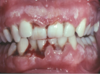
“Punched-out” interdental papillae
Necrotizing Ulcerative Gingivitis
Severe pain, oral malodor, spontaneous
hemorrhage
Necrotizing Ulcerative Gingivitis
describe Necrotizing Ulcerative Gingivitis (3)
“Punched-out” interdental papillae
Localized or diffuse gingival involvement
Severe pain, oral malodor, spontaneous
hemorrhage

Necrotizing Ulcerative Gingivitis

Necrotizing Ulcerative Gingivitis
Necrotizing Ulcerative Gingivitis
NUG - Treatment (4)
Debridement (using topical or local anesthesia)
Mild salt water rinse or chlorhexidine
Improve oral hygiene and diet
Broad spectrum antibiotic may be helpful,
particularly if systemic symptoms
aka – Herpetic Gingivostomatitis
Primary Herpes
describe symptoms of primary herpes
Acute fever, cervical lymphadenopathy, oral sores
Small ulcers often coalesce, resulting in larger
ulcers having serpentine borders
Primary Herpes

primary herpes

primary herpes
ID + tx

first 2-3 days–> acyclovir or valacyclovir (valtrex)
Symptomatic care – analgesics, antipyretics
Topical anesthetics so patient can eat and
drink – important to avoid dehydration
Popsicles can be soothing for pediatric
patients
how long does primary herpes last?
10 to 14 days
primary herpes has an approximately _____chance of developing at
least one episode of recurrent disease
Approximately 25% chance of developing at
least one episode of recurrent disease
Recurrent Herpes Two forms:
Recurrent Herpes Labialis
Recurrent Intraoral Herpes
aka – cold sore, fever blister
Recurrent Herpes Labialis
Recurrent Herpes Labialis affects where?
Affect vermilion zone or perioral skin
symptoms of Recurrent Herpes Labialis (5)
Prodromal itching, tingling, burning, erythema
followed by cluster of vesicles
what happens with Recurrent Herpes Labialis with no treatmet
With no treatment, vesicles rupture, form a
crust, and lesions heal in 7-10 days

Recurrent Herpes Labialis

Recurrent Herpes Labialis

Recurrent Herpes Labialis
Recurrent Herpes Labialis
is triggered by what
Triggered by UV light exposure or trauma
Recurrent Herpes Labialis -Treatment (4)
Avoid excess sun exposure
Sunblocks may be helpful to prevent lesion
development
Topical antiviral agents - statistically significant
decrease in healing time
Patient-initiated systemic valacyclovir seems to
have best results
*Treatment must be started within first 2-3 days of onset*
recurrent intraoral herpes occurs where?
Confined to mucosa bound to periosteum (hard
palate and attached gingiva)
describe Recurrent Intraoral Herpes
Cluster of shallow ulcers
tx for recurrent intraoral herpes
Heal in one week with no treatment

Recurrent Intraoral Herpes

Recurrent Intraoral Herpes

Recurrent Intraoral Herpes
Chronic immune-mediated disorder
Lichen planus (LP)
name two types of lichen planus
Cutaneous – may resolve in 7 – 10 yr
s • Mucosal – typically managed as chronic
condition
Cutaneous lichen planus
• Clinical features:
Purple polygonal pruritic papules with
Wickham’s striae (lacy white lines)
– Cutaneous lichen planus
• Clinical features-location
Flexor surface of wrists, lumbar region,
shins, but other locations

Cutaneous lichen planus
Oral lichen planus (OLP)
• 2 forms:
–Reticular (lacy white lines)
–Erosive (ELP)- erythematous, may
ulcerate
most common form of oral lichen planu
Reticular form
most symptomatic form of oral lichen planus
Erosive form is most symptomatic,
especially with acidic, salty or spicy foods
desribe reticular lichen planus
interlacing white lines
describe erosive lichen planus
shallow ulcers, peripheral
erythema and radiating white lines
location of oral lichen planus
Bilateral buccal mucosa, tongue, gingiva
common but any intraoral surface and
lips

lichen planus
Oral lichen planus (OLP/ELP)
• Treatment: for reticular lichen planus

usually no Tx needed
• Patient may feel “rough” areas of
hyperkeratosis, but no pain
treatment for erosive lichen planus
ELP treat with potent topical steroid
• “off label” • Systemic steroids not needed
In PV autoantibodies destroy _____ so waht happens
desmosomes
Desmosomes bond epithelial cells
together; antibodies inhibit
adherence, so a split develops in the
epithelium
describe pemphigus vulgaris
• Superficial, ragged erosions and
ulcerations
pemphigus vulgaris occurs where
Any mucosal surface
Oral lesions “first to show, last to go”
Pemphigus vulgaris (PV)
• In other words – the oral lesions
often are the initial manifestation
of the disease and the most
difficult to resolve with treatment
+ Nikolsky sign
Pemphigus vulgaris (PV)

Pemphigus vulgaris (PV)

Pemphigus vulgaris (PV)

Pemphigus vulgaris (PV)
Pemphigus vulgaris (PV)
• Treatment:
–Systemic corticosteroids, often with
azathioprine or other steroid-sparing agents
–Topical corticosteroids have little effect
Also known as cicatricial (scarring) pemphigoid
Mucous membrane pemphigoid (MMP)
Resembles PV due to blister formation
Mucous membrane pemphigoid (MMP)
Mucous membrane pemphigoid (MMP)
• linical features (con’t):
occurs where
–Any mucosal surface, occasionally affects
skin
scarring of mucous membrane pemphigoid occurs where
–Scarring
• Skin • Symblepheron (conjunctiva) • Scarring on oral mucosa rare
May see intact intraoral blisters
Mucous membrane pemphigoid (MMP)

Mucous membrane pemphigoid (MMP)

Mucous membrane pemphigoid (MMP)

Mucous membrane pemphigoid (MMP)

Mucous membrane pemphigoid (MMP)
Mucous membrane pemphigoid (MMP)
• Most significant aspect of this condition is
ocular involvement of symblepheron

Mucous membrane pemphigoid (MMP)
Mucous membrane pemphigoid (MMP)
• Treatment:
–Depends on extent of involvement
• Oral lesions alone - topical steroids,
tetracycline/niacinamide or dapsone
may be sufficient • Frequent dental prophylaxis, q 3-4
mos.
–Refer patient to ophthalmologist for
exam and follow-up
• If ocular involvement, systemic
immunosuppressive therapy indicated
Pruritus early symptom, followed by the
development of multiple, tense bullae, blisters on normal or erythematous skin
Bullous pemphigoid (BP)

Bullous pemphigoid (BP)
Bullous pemphigoid (BP)
• Treatment:
–Management similar to cicatricial
pemphigoid, but most BP cases resolve spontaneously in 1-2 years
