15 - Shoulder Joint Flashcards

1
Q

Label the following diagram.

A
2
Q

What are the three different joints of the scapula?

A
  • Glenohumeral (shoulder)
  • Acromioclavicular
  • Scapulothoracic
3
Q

What muscles insert and originate from the scapula coracoid process?

A

Insert: Pec minor and coracobrachialis

Origin: Short head biceps brachii

4
Q

What are the important bony landmarks of the lateral surface of the scapula?

A

- Glenoid fossa: head of humerus articulates here with glenoid lavbrum

  • Supraglenoid tubercle: site of origin of long head of biceps

- Infraglenoid tubercle: site of origin of long head triceps

5
Q

Where is the anatomical neck of the humerus?

A

Between the head of the humerus and the tubercles

6
Q

Label the different bony landmarks of the humerus.

A
7
Q

How do fractured scapulas occur and what is the best way to treat them?

A
  • Rare

- Severe chest trauma, e.g high speed road collisions, crushing injuries

- Tone of muscles surrounding holds fragments in place whilst healing occurs so no fixation needed

8
Q

Label this AP x-ray of the shoulder joint.

A
9
Q

What factors are responsible for the mobility and stability of the shoulder joint?

A

Mobility: (makes unstable)

  • Shallow glenoid fossa
  • Lax capsule
  • Disproportion of articular surfaces

Stability:

  • Muscles of rotator and others
  • Ligaments
  • Capsule
10
Q

What is the function of the clavicle and where does it attach to?

A
  • Attach upper limb to trunk as part of shoulder girdle
  • Protect underlying neurovascular to upper limb
  • Transmit force from upper limb to axial skeleton
11
Q

Label the important bony landmarks of the clavicle and state what each end articulates with.

A
  • Long S bone
  • Medial aspect convex anteriorly, lateral is concave
12
Q

What is the coracoclavicular ligament?

A

TWO PARTS - suspending weight of upper limb from clavicle

- Conoid tubercle: conoid ligament, medial coracoclavicular ligament

- Trapezoid line: trapezoid ligament, lateral coracoclavicular ligament

13
Q

What muscles and ligaments originate or insert onto the clavicle?

A
  • Deltoid
  • Trapezius
  • Subclavis
  • Pec maior
  • Sternocleidomastoid
  • Sternohyoid
14
Q

What type of joint is the acromioclavicular joint and what makes it atypical?

A

- Plane type synovial

  • Palpated 2-3cm medially from tip of shoulder
  • Articular surface lined with fibrocartilage not hyaline
  • Joint cavity partially divided by articular fibrocartilage disc
15
Q

Where is the joint capsule of the acromioclavicular joint?

A
  • Loose fibrous layer that gives rise to articular disc
  • Lined internally by synovial membrane
  • Posterior aspect of joint reinforced by trapezius fibres
16
Q

What are the ligaments of the acromioclavicular joint?

A

Intrinsic: Acromioclavicular ligament superiorly

Extrinsic: Conoid and trapezoid ligament

17
Q

What movement occurs at the acromioclavicular joint?

A
  • Small degree of axial rotation and anteroposterior movement
  • Passive movement as no muscles cross it
18
Q

What type of joint is the sternoclavicular joint?

A

- Saddle type synovial

  • Manubrium of sternum, clavicle and upper medial first costal cartilage
  • Only attachment of upper limb to skeleton so strong
    • Fibrocartilage lining not hyaline and articular disc*
19
Q

Why is there an articular disc in the sternoclavicular joint?

A

Allows clavicle and manubrium to slide over each other more freeling allowing rotation in 3 axes rather than 2 like a normal saddle

20
Q

What are the possible movements of the shoulder that require the sternoclavicular joint to move too?

A
  • Elevation of shoulder joint (e.g shrugging shoulders or abducting arm over 90)
  • Depression of shoulders
  • Protraction/Retraction of shoulder
  • Rotation (e.g arm over head)
21
Q

Label this diagram of the proximal humerus and state what passes through the intertubercular sulcus?

A
  • Tendon of the long head of biceps brachii
  • Edges of the groove are know as the lips and different muscles insert onto them, see on diagram

A LADY BETWEEN TWO MAJORS

22
Q

What is the danger of fracturing the surgical neck of the humerus?

A
  • Blund trauma to shoulder from falling on outstretched hand

- Axillary nerve and circumflex humeral vessels in close proximity

  • Nerve damage results in paralysis of deltoid and teres minor so hard to abduct limb. Will also have loss of sensation of regimental badge area over deltoid
23
Q

What is being pointed to here on the humerus and what is it the landmark for?

A
  • Radial groove
  • Radial nerve and profunda brachii artery lie in this groove
24
Q

What muscles attach to the humeral shaft and where?

A

Posteriorly: Lateral and medial head of triceps, with spiral groove between

Anteriorly: Corachobrachialis, deltoid, brachioradialis, brachialis

25
Q

What defines whether a muscle is intrinsic or extrinsic in the shoulder?

A

Extrinsic: Originate from torso and insert onto bones of shoulder (humerus, clavicle, scapula)

Intrinsic: Originate from scapula and/or clavicle and insert onto the humerus

26
Q

What are the extrinsic muscles of the shoulder?

A

All on back of the trunk and organised into two layers

27
Q

What are the superficial extrinsic muscles of the shoulder, their innervation, their origin/insertion and their actions?

A
28
Q

What are the deep extrinsic muscles of the shoulder, their innervation, their origin/insertion and their actions?

A
29
Q

How do you test the trapezius muscle?

A

Ask patient to shrug shoulders whilst you push down on them

30
Q

What are the intrinsic muscles of the shoulder? (scapulohumeral group)

A
  • 4 rotator cuff muscles
  • Deltoid
  • Teres Major
31
Q

What are the intrinsic muscles of the shoulder (apart from the rotator cuff) , their innervation, their origin/insertion and their actions?

A
32
Q

What are the rotator cuff muscles of the shoulder, their innervation, their origin/insertion and their actions?

A

Give glenohumeral joint extra stability

33
Q

What are the muscles of the anterior arm, their innervation, their origin/insertion and their actions?

A
  • BBC
  • Musculocutaneous nerve penetrates C and passes between bb in lateral side of the arm
  • Continues into the forearm and becomes the lateral cutaneous nerve
34
Q

What is the bicipital aponeurosis?

A

Thick fadica close to musculotendinous junction of the biceps, forms roof of the cubital fossa and blends with deep fascia of ulnar border and anterior forearm

35
Q

What does the biceps tendon reflex test?

A
  • C6 because this is predominant for supination and elbow flexion
36
Q

Where are the different places the biceps tendon can rupture, what is the signs, and what is the treatment?

A
  • Over 50 long head may rupture near scapular origin after minimal trauma, but in weightlifters distal tendon can snap near insertion
  • Patient says heard something snap in shoulder whilst lifting
  • During flexion popeye’s sign due to unopposed contracted muscle bell of biceps
  • Usually not much weakness due to supinator muscles and brachialis

- Conservative management

37
Q

What are the posterior muscles of the arm, their innervation, their origin/insertion and their actions?

A
38
Q

What are the the paths of the three nerve branches in the upper arm in the anterior brachial plexus?

A
39
Q

What does the triceps reflex test?

A
  • C7 as responsible for elbow extension
40
Q

What is the significance of the quadrangular space and what is it bordered by?

A

Route where axillary nerve and posterior circumflex humeral artery and vein enter the posterior compartment of the arm

Superior: inferior teres minor

Inferior: superior teres major

Medial: Long tri brachii

Lateral: surgical neck of humerus

Anteriorly: subscapularis

41
Q

What is the significance of the triangular space and what are its borders?

A

Circumflex scapular vessels enter infraspinous fossa, NO NERVES

Superior: inferior teres minor

Medial: superior teres major

Lateral: long tri brachii

42
Q

What is the significance of the triangular interval and its borders?

A

Radial nerve and profunda brachii artery in base of interval as they wind around humerus in the radial groove

Superior: inferior border of teres major

Medial: long tri brachii

Lateral: shaft of humerus or lateral tri brachii

43
Q

What are the main arteries of the upper arm?

A

- Brachial when axillary past inferior border or teres major

- Profunda brachii travels with radial nerve to posterior compartment of arm and terminates in anastomoses in elbow

- Brachial travels in anterior compartment and goes under brachialis where it bifurcates in cubital fossa. Travels with median nerve lateral to medial

44
Q

What is the blood supply to the anterior and posterior compartment of the arm?

A

Anterior: brachial artery

Posterior: profunda brachii

45
Q

What are the veins of the anterior arm?

A
46
Q

Where is the capsule of the glenohumoral joint?

A
  • Glenoid labrum and fossa to anatomical neck of humerus
  • Dips down medially to surfical neck
  • Laxity for full abduction
47
Q

What does the anatomical neck mark the site of?

A
  • Articular capsule
  • Epiphyseal growth plate in children
48
Q

What is the issue with a fracture of the surgical neck of the humerus?

A
  • Axillary nerve damage
  • Posterior circumflex humeral artery damage
49
Q

What is the weakest point of the glenohumoral joint and why?

A
  • Inferior, no reenforcement
  • Only depth of glenoid labrum reducing risk of dislocation
50
Q

What are the intracapsular ligaments of the glenohumoral joint?

A

- Superior

- Middle

- Inferior

From glenoid labrum to humerus to reinforce capsule anteriorly and prevent anterior dislocation

51
Q

What are the extracapsular ligaments of the glenohumoral joint?

A
  • Coracoacromial (allows supraspinatous tendon to run through)
  • Coracohumoral (base of coracoid)
  • Transverse humoral
52
Q

What are the main dynamic stabilisers of the shoulder joint and their actions?

A

Insertions of the rotator cuff muscles blend to form cuff which fuses with joint cavity strengthening it

53
Q

What are the two bursa in the shoulder joint?

A

- Subacromial bursa: under acromion to reduce friction as supraspinatus tendon passes under structures

- Subscapular bursa: between tendon of subscapularis and neck of scapula to protect tendon as it passes inferior to root of coracoid

54
Q

What structures pass through the coraco-acromial arch and what does it prevent from happening?

A
  • Subacromial bursa, long head of biceps, supraspinatus tendon, joint capsule
  • Prevents upward displacement of the humerus
  • Impingement can occur leading to irritation
55
Q

What are all the stabilisers, dynamic and static, of the shoulder joint?

A
56
Q

What muscles cause abduction of the shoulder joint?

A
57
Q

What muscles cause adduction of the shoulder?

A
58
Q

What muscles cause flexion of the shoulder?

A
59
Q

What causes extension of the shoulder joint?

A
60
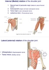
Q

What causes medial and lateral rotation of the shoulder joint?

A
61
Q

What is the nerve supply to the shoulder?

A

ALL BRANCHES OF PLEXUS WITH C5 AND C6

C5 dermatome overlies shoulder

62
Q

What is the blood supply to the shoulder?

A
  • Axillary passes behind pec minor at at surgical neck gives off anterior and posterior circumflex humeral arteries
  • Arterial anastomosis around margin of scapula. Subscapular artery (3rd branch of axillary) with suprascapular and transverse cervical
63
Q

When does the subclavian artery become the axillary artery and the axillary artery become the brachial artery?

A