13b - Clinical Correlations of Hypoxemia Flashcards
What are the factors that affect the flux of gas?
Fick’s law: Flux of gas is related to the concentration present at steady state
**Gas moves from regions of high concentration to those of low concentration with a magnitude directly proportional to the concentration gradient

What are 4 clinical causes of pulmonary diffusion defects?
Both acute and chronic conditions:
- thickening or destruction of the interstitial space, alveolar wall, or capillary
- pulmonary edema
- interstitial lung disease (sarcoid, idiopathic pulmonary fibrosis, ARDS)
- emphysema
What do you see on this xray?

Trick question: this is a normal xray (this makes a diffusion defect less likely)
A patient has the following vitals:
pH 7.45, pCO2 25, pO2 70, HCO3- 24, 88% O2 saturation
What is her A-a gradient?
A-a gradient= 150 - (5/4)(pCO2) - (pO2)
150 - (5/4)(25) - (70) = 49
**This A-a is elevated!
What are 3 possible differential diagnoses with an elevated A-a gradient?
- diffusion defect
- V/Q mismatch
- shunting
What is an ideal V/Q ratio?
V/Q= 1 where ventilation and perfusion are perfectly matched
What is an explanation for an area of persusion with low ventilation?
Pulmonary shunt
What is an explanation for an area of ventilation with low perfusion?
Dead space
What are 5 ventilation problems that would decreased the V/Q ratio?
decreased ventilation:
- asthma
- COPD
- pulmonry edema
- pleural effusions
- mucous plugging *barf sound*
What are 2 perfusion problems that would decreased the V/Q ratio?
increased perfusion:
- hepatopulmonary syndrome
- anatomic shunt
What are 3 reasons the V/Q ratio would be increased?
perfusion decreased or ventilation increased:
- PE (most common reason)
- hyperventilation (rarely pathologic)
- dead space
Describe hepatopulmonary syndrome
- normal V, increased Q (decreased V/Q)
- shortness of breath and hypoxemia caused by vasodilation in the lungs of patients with liver disease
- due to arteriovenous malformations (AVMs)
Decribe an intrapulmonary shunt
- normal Q, decreased V
- alveoli are perfused but no ventilation occurs
- main cause of hypoxemia in pulmonary edema and conditions where the lungs become consolidated (e.g. pneumonia)

Describe an anatomic shunt
- normal Q, decreased V
- bronchial arteries and coronary arteries return blood to circulation without passing by the alveoli to participate in gas exchange
- usually accounts for less than 3% of the total circulation
What components of the respiratory system comprise the physiologic dead space?
- mouth
- pharynx
- larynx
- trachea
- conducting airways
Describe anatomic dead space
- areas of the respiratory system that do not participate in gas exchange
- typically ~150 ml
Describe alveolar dead space
- not all alveolar units are as efficient in exchanging gas as they should be
- in a normal, healthy person, alveolar dead space is minimal
What does this V/Q scan show?

- pulmonary embolism
- compare the ventilation (left) and perfusion (right) scans, if they don’t show the same defects then there is V/Q mismatch
What does this V/Q scan show?

Trick question! It’s normal!
If you place a patient with a PE on 100% O2 what would you expect in regards to the arterial oxygen level?
Increased arterial oxygen! You CAN overcome V/Q mismatching with 100% FIO2 (it won’t affect a shunt however)
What are 4 causes of systemic shunts?
- Genetic conditions (Hereditary hemmorhagic telangiectasisa/HHT, VonHippel Lindau/VHL)
- Right to left cardiac blood flow (ASD, VSD, congenital)
- Pulmonary arteriovenous malformations (AVMs)
- Systemic AVMs (liver, kidney, spleen, GI, etc)
What is the Berggren Shunt Equation?
Used to compare oxygenation of a patient on room air versus 100% O2 (pulmonary shunt fraction= Qs/Qt)

How can you analyze an intracardiac shunt? Extracardiac?
- Intracardiac
- “Bubble study”
- Extracardiac
- Nuclear medicine scan
- PFT lab/Berggren shunt equation
How can you use a bubble study to locate a shunt?
- When bubbles return to left side of the heart WITHIN 4 cardiac cycles= intracardiac shunt
- When bubbles return to left side of the heart in MORE THAN 4 cardiac cycles= extracardiac shunt (e.g. hepatopulmonary shunt)
What factor has the largest effect on the oxygen content of arterial blood? What is the equation for CaO2?
Hemoglobin (Hgb) has the most effect:
CaO2= (Hgb x 1.34) x (sPO2) + (PaO2 x 0.003)
sPO2= oxygen saturation
PaO= dissolved oxygen
**Normally, CaO2= 20 g/dl
How can you calculate oxygen delivery?
Delivery (DaO2) = CaO2 x CO
CaO2= oxygen content
CO= cardiac output
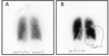
What does this xray show?

Fluid in the left lung: V/Q mismatch
What happens to a patient’s oxygen saturation following the removal of a mucous plug?
Decrease for a short time, then gradually increase to normal (due to hypoxic pulmonary vasoconstriction; diverting blood to lung areas being properly ventilated)
Describe hypoxic pulmonary vasoconstriction
- onset in seconds in response to hypoxia, max intensity in minutes
- reversible when O2 levels restored
- mitochondria sense O2 reduction and trigger response through redox sensing (avoids “wasting perfusion”)
- can be affected by volatile anesthetics, vasodilators, hypothermia, and CCBs
What endothelium-derived products increase vasoconstriction?
Thromboxane and endothelin
What endothelium-derived products inhibit vasoconstriction?
Nitric oxide and prostacyclin
