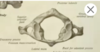
1. Skull Vault and cervical spine Flashcards

1
Q

skull vault a.k.a

A

calvaria

roof of cranial cavity

2
Q

norma verticalis

A

view of skull from above

3
Q

norma frontalis

A

view of skull from front

4
Q

norma lateralis

A

view of skull from side

5
Q

normal occipitaris

A

view of skull from behind

6
Q

frontal eminence

A

left and right sides (yellow)

most prominent part of upper forehead

similar curvature to rest of bone - more likely male

7
Q

frontal bone in young child

A

young child (2-3 years) is in 2 halves which fuse together by school age

8% have 2 halves for life

metopic suture in between

8
Q

parietal bones

A

over the parietal lobes of brain (yellow)

right and left

9
Q

parietal eminence

A

sides of skull (orange)

curvature similar to rest of skull - more likely male

more markedly curved - more likely female

10
Q

sutures

A

fibrous joints join bone of skull together

unossidied parts of membrane

assisting parts of membrane

serrated interlocking pattern

11
Q

coronal suture

A

from side to side

separates frontal bone from 2 parietal bones

12
Q

sagittal suture

A

front to back - like sagittal plane

midline of coronal suture to occiptal bone

13
Q

anterior fontanelle

A

child of 18-24 months has a diamond shaped opening called anterior fontanelle

closes completely at 2 years

metopic suture (between 2 halves of frontal bone) meets coronal and sagittal sutures

soft point

14
Q

ossification of skull

A

Bones of skull form a membrane in embryo

  • Intramembranous ossification

Membrane forms from mesenchyme

  • Mesenchymal differentiate into osteoblasts and lay down the bone

No cartilage precursor in vault of skul

15
Q

parietal foramen

A

Carries vein from veins in skull to veins inside head

Venous communication between inside and outside of cranial cavity

16
Q

lambdoid suture

A

joins sagittal suture

Upside down V

Like Greek letter Lambda

17
Q

sutural/Wormian bones

A

islands of bones seen in sutures

18
Q

sutures use in fetal life

A

Grow as new bone being laid on outside and resorbed at inside

  • Can occur at sutures

Thin bones at time of birth, sutures allow little movement of skull so baby’s head can pass through birth canal

19
Q

occipital bone

A

at rear of skull

Lambdoid suture separates parietal and occipital

Part in vault of skull

  • Squamous part of occipital bone

Part in base of skull

  • Basilar part

below occipital protuberence

20
Q

external occipital protuberence

A

most posterior part of skull

muscles of neck attach below that

21
Q

frontal bone

A

Forehead and roof

Gabela – midline front above bridge of nose

Coronal suture separates from parietal bones

22
Q

temporal bone

A

Passage of time – hear goes grey at this area first

Squamous part

  • Flat piece on side of vault of skull

Zygomatic process

  • Comes forward – meets zygomatic bone at suture
  • Zygomaticotemporal suture

Mastoid part

  • Rear on outside

Mastoid process - behind ear

  • Helps differentiate sex – males larger than females in general
  • Forms at age of 2

External acoustic metauts with bone on outside tempanic plate

  • Styloid process - Long bone process done to mandible

Petrymastoid parts

  • Dense bone inside skull
23
Q

sphenoid bones

A

butterfly shaped

greater and lesser wings

lies on floor of cranial cavity across midline

extends to side of skull and contributes to normal lateralis - greater wing

24
Q

pterion

A

sutures join in H shape

Means wing, like Mercury God

Thin area of skull – can be easily broken

  • Middle meningeal artery run in grove below this

Extradural haemorrhage risk

25
temporal process of zygmatic bone
26
zygomatic process of temporal bone
27
tympanic plate
28
superior and inferior temporal lines
faint For temporalis muscles attachment to inferior line Dense fascia covers the temporalis muscle to superior line Lines downwards is temporal fossa where temporalis muscle lies
29
zygomaticofacial foramen
sensual nerve onto zygoma
30
bregma
point where coronal and sagittal sutures meet
31
maxilla
upper jaw right and left sieds joined by suture contributes to part of nasal aperture and lower mesial side to oribital margins and floor of orbit
32
right and left nasal bones
small thin bones attached to frontal bones maxilla and each other can be easily broken
33
nasal aperture
upper edges formed by nasal bones pear shaped
34
zygomatic bones
prominent part of cheek lateral wall of orbit can feel frontal zygomaticofacial suture on palate
35
mandible in fetus
in 2 halves - joined by fibrous joint so single by birth
36
bones inside eye
* sphenoid bone * lacrimal bone * ethmoid bones not part of normal frontalis
37
supra orbital foramen
Notch or hole Supraorbital nerve - Sensory nerve to forehead, branch of CNV3 ophthalmic * Sensation and blood supply in parallel to forehead
38
infraoribital foramen
infraorbital nerve sensory nerve to front of cheek, side of nose, lower eyelid and upper lip and gingivae, continuation of CNV2 maxillary division
39
mental foramen
Coming out of mandible Inferior alveolar nerve – CNV3 mandibular division – runs in bone and comes out as mental nerve at premolar teeth region Sensory nerve to skin of chin, lower lip and gingiva
40
state of tooth loss
Healed sockets antemortem -mandible Open socket postmortem – maxilla
41
punch on front of face can cause
bruise to infraoribital nerve as large foramen numbness to fronot of face
42
typical cervical vertebrae
C3-6
43
body of typical cervical vertebrae
Not flat Curved from side to side * Sides rise higher than central
44
vertebrae foramen
vertical cervical cord surrounded by meninges outside of meninges is fat and venous plexus
45
vertebra arch
behind foramen made of * pedicle * lamina
46
spinous process
posterior to vertebra cervical splits into 2 - bifid
47
transverse process
side of vertebra Has hole – foramen transversarium * Carries vertebral artery in C1 to C6 * Not C7 In front of hole – costal element * Equivalent of rib at lower levels (reduced in Cervical section)
48
intervertebral discs
- Fibrous cartilage Classified as secondary fibrocartilage joint/symphysis - Bone, fibrocartilage, bone
49
what joins vertebrae together
facets Covered by cartilage * Smooth surface Superior to inferior above Synovial joint * Capsule lined by synovial membrane inside the joint * Synovial fluid Subject to diseases * Osteoarthritis * Osteophytes – osteoarthritis disease of bony projection
50
diseases in vertebrae
Osteoarthritis Osteophytes – osteoarthritis disease of bony projection * pain due to osteophytes at pedicle – press on nerve and radiate onto upper limb if after C5
51
how do spinal nerves leave spinal canal
by passing over the pedicle bone
52
changes in grove for nerve
gets larger as go down the spine as nerves inc in size (begin to form the brachial plexus)
53
atlas
C1 atypical no ventral body * has anterior arch of atlas (bar of bones) no spinous process wider transverse process
54
axis
c2 atypical Bony peginate * Odontoid process a.k.a Dens - Looks like canine Held up against inside of anterior arch of atlas * Ligaments pass behind that * Transverse ligament of head Rotate head * Most of movement from between the atlas and axis C2 has a large transverse process – not bifid
55
rotation of head
Most of movement from between the atlas and axis Synovial joint of pivot variety * Atlas can turn around dens Facets between atlas and axis are flat to allow rotation to take place
56
C3, 4, 5, 6
TYPICAL CERVICAL VERTEBRAE * Concave body * Tranverse process – before nerve splits into ventral and dorsal rami * Superior articular facet * Lamina * Pedicle * Bifid spinous process * Notch reflects level of vertebra – larger as down
57
C7
atypical Obvious large spinous process * Not bifid * Like thoracic spinous process Vertebral Artery doesn’t pass through foramen transversarium (has one though)
58
where is upper cervical spine
directly behind oral cavity
59
what is this
dens of axis C2 helpful to ID
60
what is this
flat articular facets of C2
61
what is this
body of C2
62
what is this
C3
63
what is this
lateral parts of axis
64
atalantoaxial joint
Cartilage covering bones creates joint space No evidence of arthritis here
65
what is this
transverse process of atlas wide and feel behind lower of ramus of mandible
66
what is this on lateral x ray of cervical spine
body of axis
67
what is this on lateral x ray of cervical spine
anterior arch of atlas resting against dens to allow pivot rotation
68
what is this on lateral x ray of cervical spine
posteiror arch of atlas not a spinous process
69
what is this on lateral x ray of cervical spine
C6
70
what is this
intervertebral foramen opening between 2 vertebrae spinal nerve through
71
cervical nerve exits points C1
over atlas
72
cervical nerve exits points C2
between atlas and axis
73
cervical nerve exits points C3
between C2 and C3
74
cervical nerve exits points C4
between C3 and C4
75
cervical nerve exits points C5
between C4 and C5
76
cervical nerve exits points C6
between C5 and C6
77
cervical nerve exit points C7
between C6 and C7
78
cervical nerve exit points C8
between C7 and T1
79
cervical nerve exit points T1
T1 comes out below pedical of T1 and so on
80
cervical lordosis
vertebral coloum is curved so convex forwards
81
carnium
top part of bony box for brain
82
organs in front of cervical spine
* Air passage – nasal and oral passages * Oesophagus * Thyroid gland Organs tend to be in midline and out to the side is blood vessels and lymphatics (vascular arrangements) along with Cranial Nerves
83
glasgow coma scale
how deeply unconscious the pt is Response to pain * Press on supra-orbital notch Third along orbit from midline – dull drilling pain
84
zygomatic buttress/process of maxilla
towards the zygomatic bone - Obvious in dental radiology
85
bragma
the coronal suture and sagittal suture meet
86
vertex
highest point of skull
87
emissary vein exits
parietal foramen venous connection bewteen inside and outside skull infection in skull, potential route of spread into cranial cavity
88
most curved partietal eminence likely
female
89
simiarl curvature for parietal and frontal eminence likely
male
90
locations of squamous and basilar points of occipital bone
squamous at back basilar towards foramen magnum
91
external occiptal protuberence
bulge at most posterior of head inion: middle point of the external occiptal protuberence
92
inion
middle point of external occipital protuberence
93
zygomatic arch
zygomatic process of temporal and temporal process of zygomatic bone - Can fracture
94
what holds the dens in place
ligaments – loosening of ligament dangerous, hold tightly but allow dens to swivel (pivot joint)
95
what is this
dens
96
what is this
C4
97
C1-3 location in relation to front of body
C1 and C2 behind mouth C3 at hyoid bone
98
fratcure of front of cranial cavity dabbing fluid at nose
could be draining cerebral spinal fluid, olfactory nerves can be broken, escape into nasal cavity – sample and send to biochemistry Mucous sticky fluid – carbohydrates/glycoproteins (unlike CSF – more water based) Frontal sinus exposure potential, floor of anterior cranial cavity breached as well as dura – possible ingress of bacteria into meninges then meningitis risk