1 - Neuro-Ophthalmic Anatomy Flashcards
1
Q
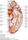
Skull Base Anatomy and Foramina
A
- Standing Room Only — Foramina of Trigeminal Nerve branches
- S for Superior Orbital Fissure —- V1 Ophthalmic; Lacrimal, Frontal, Nasociliary
- R for Rotundum — V2 Maxillary Nerve
- O for Ovale — V3 Mandibular Nerve, Lesser Petrosal Nerve branch of CN9 (Glossopharyngeal) which innervates parotid gland
- Foramen Spinosum — Middle Meningeal Artery branch of ECA
- Foramen Lacerum — Greater Petrosal Nerve branch of CN7 which innervates lacrimal gland
- Carotid Canal — ICA

2
Q
Sphenoid Bone
A

3
Q
Sella Turcica
A

4
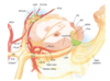
Q
Superior Orbital Fissure
A
- CN3 inferior and superior division
- CN4
- CN6
- V1 (Ophthalmic Nerve) of Trigeminal Nerve. Includes Lacrimal, Frontal, and Nasociliary of V1
- Sympathetic fibers

5
Q
Bones of Orbit
A
- 7 Bones
- Maxillary
- Zygomatic
- Frontal
- Lacrimal
- Sphenoid
- Palatine
- Ethmoidal

6
Q
Orbital Roof
A
- Frontal + Lesser Wing of Sphenoid
- Medially frontal bone forms roof of ethmoidal sinus
7
Q
Supraorbital Nerve
A
- From Frontal Nerve
- Travels through Supraorbital Notch

8
Q
Supratrochlear Nerve
A
- From Frontal Nerve
2.

9
Q
Sensory area of Supraorbital and Supratrochlear Nerves
A

10
Q
Sensory area of Supraorbital and Supratrochlear Nerves
A

11
Q
Lateral Orbital Wall
A
- Zygomatic + Greater Wing of Sphenoid Bone
- Junction between lateral orbital wall and roof = frontosphenoid + frontozygomatic sutures
- Separates orbit from middle cranial fossa
- At 90° angle from each other

12
Q
Orbital Floor
A
- Zygomatic + Maxillary + Palatine
- IO originates laterally to NLD

13
Q
Infraorbital Groove
A

14
Q
Medial Orbital Wall
A
- Sphenoid, Maxillary, Ethmoid, Lacrimal
- Largest component = Ethmoid
- Lacrimal Sac Fossa = Fromed by Maxillary and Lacrimal bones
- Lacrimal bone divided by posterior lacrimal crest
- Anterior lacrimal crest = Maxillary bone

15
Q
Orbital Dimensions
A
- 45mm wide
- 35mm height max
- Volume = 30cm3
- Medial Wall from Rim to Optic Canal = 40mm
- Medial Walls parallel
- Lateral Walls form 90° angle
16
Q
Paranasal Sinuses
A

17
Q
Importance of frontoethmoidal suture?
A
- Marks inferior boundary of anterior cranial fossa
18
Q
Location of Sphenoid Sinus
A
- Forms medial wall of optic canal
- Route for decompression of optic chiasm

19
Q
Sphenopalatine Ganglion AKA Pterygopalatine ganglion
A
- Underlies Apex of Orbit

20
Q
Optic Canal Contents
A
- Optic Nerve
- Ophthalmic Artery
- Some sympathetic fibers
21
Q
Pterygopalatine Fossa
A

22
Q
Pterygopalatine Fossa
A
- Orbit connected to pterygopalatine fossa via inferior orbital fissure

23
Q
Contents of Inferior Orbital Fissure?
A
- Zygomatic Nerve and Infraorbital Nerve Branch of Maxillary (V2) which is Branch of Trigeminal
- Inferior ophthalmic vein
- Infraorbital artery and vein
- Branches from pterygopalatine ganglion
- Parasympathetic fibers

24
Q
Arterial Supply
A
- CCA originate from inominate artery from R
- CCA originate from aorta on L
- Vertebral arteries join near pontomedullary junction to form basilar artery
- Basilar artery ascends along anterior surface of pons and terminates into 2 posterior cerebral arteries at level of midbrain
- CCA divides into ECA and ICA at angle of jaw
- ECA supplies blood to face via facial artery
- Posterior scalp = occiptal artery
- Anterior scalp = superficial temporal artery

25
Collateral Anatomy

26
ICA Pathway

27
ICA Pathway

28
CRA enters ON where?
10-12 mm posterior to globe
29
Ophthalmic Artery and Branches

30
Ophthalmic Artery and Branches

31
Maxillary Artery and Branches

32
Muscular Arteries do what?
1. Superior Branch --- Superior Rectus, Superior Oblique
2. Inferior Branch --- Medial Rectus, Inferior Rectus
33
Lacrimal artery
1. From Ophthalmic artery
2. Provides Lateral Rectus
3. Gives off anterior ciliary artery which supplies blood flow to anterior ciliary body and anastomoses with long posterior ciliary artery to form greater arterial circle

34
Short Posterior Ciliary Arteries
1. 10-20 small branches --- supply ONH and posterior choroid, RPE, photoreceptors, outer 1/3 of retina
2. 30% of people branch of posterior ciliary artery called cilioretinal artery directly supplies portion of inner retina which may protect macula in case of CRAO
3. Circle of Zinn-Haller --- supplied by anastomoses of 4 SPCAs

35
Collateral of Eyelid
1. Transverse facial and frontal branch of superficial temporal branch of ECA

36
Circle of Willis

37
Anterior Communicating Artery
1. ACA crosses over optic nerve and joins opposite ACA via ACoA thus aneurysm of ACoA may cause visual defect
2. Small branches of from proximal ACA and ACoA supply intracranial optic nerves and chiasm
3. Distal ACA occlusion may cause disruption of premotor areas of frontal lobe responsible for initiating saccades with difficulty initiating saccades to contralateral side

38
AChoa supplies what?
1. Anterior choroidal artery supplies LGN
39
Middle Cerebral Artery (MCA)
1. Branches supply optic radiations
2. Terminal branches also supply occiptal tip representing macula which is resposnsible for macular sparing in PCA or calcarine artery occlusion
3. MCA occlusion may disrupt visually guided pursuit movements

40
MCA distribution

41
Posterior Circulation

42
Posterior Circulation
1. VA or PICA occlusion causes lateral medullary syndroma aka Wallenberg syndrome

43
Basilar Artery
1. Small perforators arise and supply PPRF, MLF, medially located nuclei of CN3, 4, 6.
2. SCA supplies CN3 and its fascicles
44
PCA
1. CN3 exits between SCA and PCA
2. PCoA parallels course of CN3
3. Calcarine branch of PCA supplies visual cortex

45
Venous drainage
1. Central Retinal Vein from retinal veins
2. Vortex Veins from choroidal veins
3. Episcleral Venous Plexus from anterior uvea + aqueous
3 primary venous drainage pathways empty mainly into SOV
46
Optociliary shunt vessels?
1. Shunt connecting retinal veins to choroidal veins usually seen in CRVO or ON meningioma
47
Blind Spot
1. Due to absence of retinal receptors over ONH
2. Located 17 degrees from fovea measuring 5 degrees by 7 degrees
3. Fovea ~ 1.5mm or 1dd. Located 4mm from and 0.8mm lower than ONH
48
Intrinsically photosensitive retinal ganglion cells (ipRGCs)
1. Subset of RGCs that serve primarily nonvisual light-dependent function such as pupillary light reflex
49
Ratio of photoreceptor cells to RGCs
1. 1000:1 in periphery
2. Lowest ratio in fovea (RGC may receive signal from 1 cone cell)
3. 69% of RGCs within central 30 degrees thus foveal bipolar cells and RGCs displaced radially from fovea and receive input from long cone axons that make up Henle Layer
50
Optic Nerve contains how many RGCs?
1. 1-1.2M RGCs
51
ON posterior to sclera
1. Acquires myelin from oligodendrocytes which increases diameter
52
Optic Nerve regions
1. Intraorbital 30mm allowing globe rotation
2. Blood supply mostly from pial branches of Ophthalmic artery
3. Peripheral retinal receptors --- found peripherally
4. Papillomacular bundle --- travels temporally
5. Optic Canal 8-10mm long and 5-7mm wide
6. Intracanlicular ON susceptible to damage from sinus surgery and processes that cause bone thickening such as fibrous dysplasia, intraosseus meningioma
7. Intracranial 8-12mm. May be impinged by lesions of sella

53
Optic Chiasm
1. Supplied by proximal ACA and ACoA
2. Directly above sella
3. 53% nasal retina fibers cross
4. Wilbrand knee = anterior loop of fibers into contralateral optic nerve
54
Optic Tract
1. Just prior to LGN fibers involved in pupillary pathways exit to pretectal nuclei

55
LGN
1. 6 levels
1. 4 superior levels = P cell axons which are ganglion cells with smaller receptive fields responsible for spatial resolution and color perception
2. 2 inferior levels = M cell fibers, which are ganglion cells with larger receptive fields and more sensitive to motion detection
3. Contralateral eye 1,4,6 layers
4. Ipsilateral eye 2,3,5 layers
56
Optic Radiations

57
Primary Visual Cortex
1. AKA V1, striate cortex
2. Optic radiations terminate in 4th layer of 6 layers of primary visual cortex
3. 50-60% of cortex responds to activity within central 10 degrees
4. 80% of cortex devoted to macula = central 30 degrees

58
Parallel visual processing pathways
1. V2
2. V3 = Visual integration
3. V4 = Color
4. V5 = movement and direction of moving stimuli, origin of pursuit movements
5. Occiptotemporal pathway --- What pathway
6. Occipitoparietal pathway --- Where pathway

59
Saccadic system
1. From 2 parallel interconnected descending pathways
1. Posterior parietal cortex = visually reflexive
2. Frontal Eye Field = memory-guided, visually guided and volitional saccades
2. FEF controls gaze to opposite side thus stroke on R side cause deviation to R side

60
Smooth Pursuit System
1. Originates in V5 aka MT
2. Doubly decussates thus ipsilateral system

61
Important structures involved in eye movements
1. Rostral Interstitial nucleus of MLF = excitatory neurons that generate vertical and torsional saccades. Within midbrain
2. Interstitial nucleus of Cajal = inhibitory burst neurons for vertical saccades and neural integrator for vertical and torsional gaze
3. Nucleus prepositus hypoglossi = neural integrator for horizontal gaze
4. PPRF = excitatory burst neurons that generate horizontal saccades and inhibitory burst neurons for horizontal saccades
5. CN6 = horizontal smooth-pursuit eye movements
6. In general, midbrain for vertical eye movements and pons for horizontal eye movements

62
Vestibular-Ocular System

63
Ocular Motor CN
1. EOMs receive innervation 1/3 of distance from apex except for IO
2. IO receives innervation at midpoint
3. EOMs receive innervation on inside surface except SO which receives CN4 input on upper outer surface
64
CN6 Path
1. Originates in dorsal caudal Pons beneath 4th ventricle
2. Nucleus surrounded by looping fibers of CN7 and adjacent to PPRF
3. Exits brainstem and runs rostrally (superiorly) within subarachnoid space on surface of clivus. Pierces dura and 1cm below petrous apex and travels beneath petroclinoid ligament and enters Dorello canal
4. Once extradural, CN6 within Cavernous Sinus parallel to Ophthalmic Nerve V1
5. For short segment anteriorly joined with sympathetic chain within anterior cavernous sinus
6. Travels through SOF and through annulus of Zinn and enters medial surface of LR

65
CN6 brainstem location

66
CN6 Path

67
CN6 and Cavernous Sinus

68
CN4 Path
1. Nucleus within dorsal caudal midbrain
2. Crosses around aqueduct
3. Exits on dorsal surface of brainstem
4. Longest unprotected intracranial course
5. Enters lateral Cavernous Sinus
6. Enters SOF outside and superior to annulus of Zinn

69
CN3
1. Nucleus in dorsal midbrain
2. Exit brainstem
1. SR fibers cross first before exiting
3. In subarachnoid space passes between SCA and PCA
4. Pupillary fibers travel on dorsomedial surface of nerve
5. Superior division (SR, LPS) and Inferior division (MR, IR, IO, pupillary sphincter & ciliary body muscles via ciliary ganglion) divide at level of anterior cavernous sinus/SOF
6. Within annulus of Zinn
7. Inferior division sends parasympathetic fibers to ciliary ganglion 10mm anterior to annulus of Zinn and lateral to optic nerve. After synapsing in ganglion, accompany IO fibers
8. 9-10x more accomodation fibers than pupillary fibers

70
CN3
1. Lesion to CN3 nucleus will cause bilateral ptosis as SR nuclei very close and damage to one will damage other one

71
Oblique EOMs
1. Oblique muscles insert posterior lateral aspect of globe
2. Oblique muscles form 51 degree angle with visual axis in primary position
3. When globe adducted 51 degrees then act as elevators/depressors
4. IO originates anteromedial periorbita near posterior margin of lacrimal fosss
5. IO primary action (turning) excyclotorsion, secondary action (up/down) elevation, tertiary action (in/out) abduction
6. SO primary action incyclotorsion, secondary action depression, tertiary action abduction

72
Oblique EOMs

73
Rectii EOMs
1. Rectii insert medial obliquely oriented out
2. Rectii form 23 degree angle with visual axis in primary position
3. When eye abducted 23 degrees rectii act in elevation/depression
4. SR primary action elevation, secondary action (turning) intorsion, tertiary action adduction
5. MR primary action elevation, secondary action extorsion, tertiary action adduction

74
CN5
1. Nuclear complex extends from midbrain to cervical spinal cord
2. 3 sensory nuclei and 1 motor nuclei
1. Main sensory nucleus --- light touch from skin of face and mucous membranes
2. Mesencephalic nucleus --- proprioception and deep sensation of facial muscles
3. Spinal nucleus --- extends to C4, pain and temperature information
4. Motor nucleus --- mastication muscles, tensor tympani, tensor veli palatani, digastric muscle
3. Sensory nuclei project to thalamus
4. Trigeminal ganglion located in floor or middle cranial fossa = Meckel Cave

75
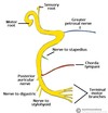
CN5 - V1
1. Runs within lateral wall of cavernous sinus
2. Divides into lacrimal, frontal and nasociliary as it approaches SOF
3. Lacrimal and Frontal enter apex of orbit outside of annulus of Zinn
4. Frontal nerve divides into supraorbital and supratrochlear --- forehead, frontal sinus, upper eyelid, conjunctiva
5. Lacrimal nerve --- lacrimal gland
6. Nasociliary branch enters annulus of Zinn, runs through ciliary ganglion and innervates globe through short and long posterior ciliary nerves --- lateral wall of nose, skin of nose to nasal tip

76
CN5
1.

77
CN5 - V2
1. Nasopharynx, hard and soft palate, portions of nasal cavity, upper gums and molars, lateral face, cheek, lower eyelid, upper teeth

78
CN5 - V3
1. Via foramen ovale
2. Skin of jaw, and carries motor division of CN5 to muscles of mastication and neck

79
CN7
1. Path starts in precentral gyrus (primary motor cortex) → Upper Face receives neurons from bilateral primary motor cortex → **Lower Face receives neurons from ONLY from contralateral cortex** → CN7 nucleus also receives input from Basal Ganglia responsible for involuntary blinking.
2. Thus cortex lesion (supranuclear lesion) on 1 side will cause ONLY lower facial palsy on contralateral side because upper face receives bilateral input

80
CN7 Nuclei
1. Motor fibers originate in caudal pons
2. 3 nucleii and 2 roots (motor and sensory root)
1. Nucleus Solitarius
2. Motor Nucleus
3. Salivatory Nucleus
3. CN7 primarily Motor in function
4. After bending around CN6 nucleus, CN7 exits at cerebellopontine angle
5. CN8 + Motor root of CN7 + Nervus Intermedius (sensory and parasympathetic root of CN7) enter internal auditory meatus

81
CN7 Path from Geniculate Ganglion
1. Sensory and Motor Root Fuse as they leave internal auditory meatus to form Facial Nerve
2. Sensory cells of Nucleus Solitarius continue through geniculate ganglion. Distally continue as **chorda tympani** and with lingual nerve of V3 → taste fibers. Synapse in submandibular ganglion → salivation via Submandibular/Sublingual gland
3. Cells from Salivatory nucleus continue through geniculate ganglion via **greater petrosal nerve.** Synapse in sphenopalatine ganglion → lacrimal (lacrimal gland)
4. Motor Nerves → Nerve to stapedius, posterior auricular nerve, digastric nerve, stylohyoid nerve
5. As Facial Nerve passes through parotid gland gives terminal 5 branches
1. Temporal
2. Zygomatic
3. Buccal
4. Marginal Mandibular
5. Cervical

82
CN7 Branches
83
Sympathetic Pathway
1. Hypothalamus → anteromedial column of brainstem → intermediolateral column of C-spine → **synapse** C8-T2 ciliospinal center of Budge-Waller → ventral rami of C8, T1, T2 levels and joing paravertebral sympathetic plexus → passes close to inominate artery in R side and subclavian artery on L side just above lung apex → **synapse** in superior cervical ganglion at level of angle of jaw C2 and carotid bifurcation → 3rd order neurons continue in wall of bidurcated carotid with fibers innervating sweat glands of lower face following ECA and fibers for pupil following ICA → some fibers leave ICA and joing greater superficial nerve to form vidian nerve which parallel parasympathetics to lacrimal gland; within cavernous sinus fibers for dilator muscle join CN6 for a few mm → further anteriorly fibers join nasociliary branch of V1 → in apex fibers pass through ciliary ganglion without synapsing and along with nasociliary branch travel with long ciliary nerves to dilator muscle of pupil → fibers destined for Muller muscle travel along ophthalmic artery and frontal and lacrimal branches

84
Parasympathetic Pathway
1. Fibers contolling pupil sphincter muscles originate in Edinger-Westphal nuclei of CN3 within midbrain
2. Main input to EW from pretectal nuclei which receive input from afferent visual pathway anterior to LGN
3. Cortex, frontal lobes, hypothalamus, reticular activating system provide tonic inhibitory signals to EW
4. Parasympathetic fibers + CN3 fascicle leave CN3 nucleus and within subarachnoid space parasympathetic fibers run on medial superficial surface of CN3 → CN3 bifurcates in anterior cavernous sinus → parasympathetic fibers travel with inferior division → synapse in ciliary ganglon → travel with branch destined for inferior oblique muscle → join short posterior ciliary nerves to reach anterior segment and iris sphincter
5. Parasympathetic innervation to lacrimal gland originates in superior salivatory nucleus and receives input from CN5 ancd hypothalamus. Eventually through geniculate ganglion → sphenopalatine and submandibular ganglion. **Fibers through sphenopalatine ganglion travel through inferior orbital fissure and then with lacrimal nerve to reach lacrimal gland.**

