(01) Amyloid Flashcards
(What is amyloid?)
- Not a single substance - but a class of what?
- located intracellularly or extracellularly?
- called amyloid cause it stained the same way as starch (thanks Rudolf)
- What kind of compound is it?
- What is amyloidosis?
- insoluble abnormal protein deposits in tissue
- can be either
- mainly protein with some COH components
- disease condition where amyloid is deposited locally or systematically
(Amyloid Classification)
- What type involves more than one organ?
- What one is restricted to a single organ or tissue?
- which type is more common?
- systemic (eg liver, kidney, GIT and spleen in AA amyloidosis)
- localized (eg pancreatic islets in islet amyloidosis)
- localized

note all the little speckles in the cortex - this is glomeruli where amyloid has been deposited

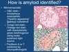
(How is amyloid identified)
(microscopically)
1-3. What are the main three stains used?

- H&E stain
- Congo Red Stain
- Thioflavin S or T
(and then read the slide)
amyloid can bend the light coming through - that is what befriegence is I guess
at EM level - all amyloidosis will look essentially the same
this is the glomerulus - a capillary loop
normally albumin and everything larger will be retained - when amyloid blocks the filtration barrier you get lots of proteins spilling out of the glomeruli
as deposits grow the lumen also gets smaller - going to ischemia when it is small enough - this will cause damage to a nephron - will get chronic renal failure once things get bad enough


(Amyloid Structure/Composition)
(Fibrils)
- what are they?
- Protein secondary strucutre is what?
- Over 20 different protein precursors have been identified - all have regions that do what?
- polymerized repepitive homologous peptide subunits
- B-pleated sheet conformation
- have a propensity to form B-pleated sheet secondary structure
HE wants to make one point
this is a B-pleated sheet
what happens when B-sheets align next to one another - H-bonds form all along the chain - this makes these structures very stable (very insoluble)

congo red happens to have a conformation that allows it to line up with grooves

much of the damage cause by amyloidosis is caused by oligomers early on in the formation of these structures
oligmers of various sorts can cause cytotoxicity - can damage cell membranes - (in diabetes and alzheimers for example) - damage is caused by the formation of the oligomers

(Significance of Amyloid)
- What is replaced by amyloid?
- Why does amyloid tend to accumulate in tissues?
3-4. Accumulation can lead to what two things (in regards to damage type)?
- functional tissue
- due to its insolubility
- physical compression of normal tissues resulting in atrophy (Eg. AA amyloid in liver sinusoids)
- disruption of normal structures and impaired function - Eg glomerular basement membrane
(pictured - glomerulus replaced with amyloid)

(Significance of Amyloids)
- Amyloid oligomers and/or fibrils may be cytotoxic and induce what and what?
2-3. What are two examples?
- cell stress responses and apoptosis
- B-cell death associated with islet amyloidosis (AIAPP) leading to type 2 DM
- neuronal death in Alzheimer’s disease (Abeta “amyloid plaques)
(pictured: upper left - cat islet (normal)) - (lower right - pancreatic islet replaced by amyloid)

(Pathogenesis of amyloidosis)
- Not completely understood by probably multiple mechanisms involved
2-4. What are the three reasons?
- increased protein production of precursor protein (Eg likely mechanism in islet amyloidosis and also involved in AA, AL (derived from immunoglobin light chains) and other forms
- Mutation to precursor protein resulting in more amyloidogenic form (usually results in increased propensity to form B-sheet structure) (eg Serine20Glutamate in human IAPP –> early onset type 2 DM)
- Abnormal processing of precursor protein to form amyloidogenic polypeptides (eg. probable role in Alzhemier’s disease amyloid formation (APP to B-protein))
AA = A(myloid)A(serum amyloid A)
AL = A(myloid)L(immuglobulin light chain)
(AA (secondary or reactive) Amyloidosis)
- Most serious systemic form in what and what? may lead to what?
- A systemic amyloidosis which occurs secondary to what?
- IL-1 –> ?
- Macrophage degradation of 104 Amino Aacid residue SAA to 76 residue amyloidogenic fragment –> ?
- can occur where?
- mammals and birds; renal and hepatic failure
- recurrent or chronic disease (chronic inflammation/infection, neoplasia)
- increased hepatic synthesis of SAA
- amyloid
- liver, kidneys, spleen, blood vessels, GI
(Canine Amyloidosis)
- What is the most serious systemic form?
- Clinical signs most often related to what with what and what?
- with severe liver involvement wha may occur?
- systemic AA (secondary/reactive) amyloidosis
- to renal glomerulus with marked proteinuria and renal failure
- hepatic failure
(can also have failure of both - depends where it is depositing)
(Canine Amyloidosis)
- Familial form of AA amyloidosis in Shar Pei dogs develops secondary to what?
- AA amyloidosis with what and what?
- Shar Pei AA amyloid sequence same as other breeds - what does this mean?
- Recently what has been linked to this condidtion?
- Shar Pei fever
- renal medullary interstital deposits (not so much in glomeruli) and chronic renal failure
- isn’t the genetic cause
- mutation in HAS2 gene
(Canine amyloidosis cont.)
- What is the most common systemic form?
- Found in what percentage of dogs over 10 years?
- derived from what?
- are there usually clinical manifestations? rarely may be associated with what?

- pulmonary vascular amyloid
- 22% of dogs
- apolipoprotein A1
- no; pulmonary hemorrhages
(pictures are of amyloid in vessels)

(Canine amyloidosis cont)
(beta-protein derived (Alzheimer’s like) cerebral amyloid)
- amyloid plaques occur in 36% of Beagles 11-3 years old and in 73% of Beagles 15-18 years old
- Neuropathology includes what and what?
- Associated with cognitive dysfunction - similar pathologically and clinically to Alzheimer’s disease
- senile plaques (amyloid) and cerebral vascular amyloidosis
(these are amyloid plaques from human alzheimer’s patient)

(Feline Amyloidosis)
(Systemic AA amyloidosis)
- how common? except for what breeds? What do they develop?
- What may occur in affected cats?
- What appears to increase its amyloidogenicity?
- uncommon; abyssinian, Siamese, and Oriental short hair cats; familial forms of systemic AA amyloidosis with hepatic (and sometimes) renal amyloidosis
- hepatic failure and rupture
- mutation in the SAA gene
(Feline Amyloidosis (cont))
(Islet Amyloidosis - a localized form of amyloidosis)
- nearly all type 2 diabetic cats have partial replacement of their islets with what?
- IAPP is a hormone produced by what and co-secreted with what?
- long standing insulin resistance –> ? –> ?
- small IAPP amyloid oligomers are most cytotoxic and may also do what?

- amyloid derived from islet amyloid polypeptide (IAPP)
- produced by pancreatic B-cells, co-secreted with insulin
- increased production of IAPP –> plus unkonwn factors
- disrupt cell membranes
(pictured - front - pancreas from diabetic cat - can see amyloid in one of the islets)
(back - closer up view)

(Bovine Amyloidosis)
- Systemic AA form
- involvement in what or what?
- Often associated with what?
- renal (proteinuria) and/or GI (severe diarrhea)
- foot abcesses and necrotizing pododermatitis (chronic inflammation/infection)
(Equine Amyloidosis)
- Systemic AA form occurs occasionally - and often in horses used for antiserum production - involvement of what two organs?
- Localized forms (AL) occur as what? or what else? both forms are of what origin?

- liver or GI
- a nodular skin form; multinodular nasopharyngeal form (difficult breathing); Ig light chain
(pictured: front - horse liver with AA amyloid - congo red stain)
(back - thinks its the spleen again but not sure…)

(Systemic AA Amyloidosis in ZEW species)
(captive cheetahs)
- frequent occurence of what form? associated with what?
- Amyloid deposits mainly in what?
- What is the main clinical condition?
- AA amyloidosis; inflammatory conditions (mainly gastritis)
- kidney (medullary interstitium, less in glomeruli) & liver
- chronic renal failure

(Birds)
- What is a major form encountered?
- High incidence in waterfowl may be related to what three things?
- Systemic AA amyloidosis
- genetic influences, chronic infection, and stress
(Gross/microscopic features of AA amyloid)
(Spleen)
- may be multifocal “sago spleen” when it involves what?
- Or diffuse in the red pulp - causes what?
- the white pulp
- generalized enlargement of the spleen
(Gross/microscopic features of AA amyloid - Liver)
- common site in what form?
- see what?
- First where, replaces what, atrophy of what?

- systemic form
- hepatomegaly, pale/yellow, heavy, friable
- space of Disse, sinusoids, hepatic cords
(pictured - liver getting amyloided)

(Gross/Microscopic features of AA amyloid - Kidney)
- Kidneys are commonly invovled in what form?
- mottled pale tan and red with prominent what?
- glomerular deposits where and where and where?
- advanced stages progress to what?
- AA systemic form
- glomeruli
- mesangium, within the capillary walls, and GBM
- obliteration of glomerulus
