NECK - vasculature & lymphatics Flashcards

1
Q

What are the borders of the ANTERIOR triangle?

(medial, lateral, superior)

A

Medial = Midline of neck

Lateral = Anterior border of sternocleidomastoid

Superior = Inferior border of mandible

2
Q

What are the 4 blood vessels found in the ANTERIOR trinagle?

(4)

A

Common carotid a.

External carotid a.

Internal carotid a.

Internal jugular v. + tributaries

3
Q

What blood vessels are found in the CAROTID triangel (sub-region of anterior)?

(4)

A

common carotid a.

external carotid a.

internal carotid a.

Internal jugular v.

4
Q

Trace the ascent of the R. common carotid a.:

A

brachiocephalic trunk (posterior to R. sternoclavicular joint) –> R. common carotid a.

  • Ascent through neck w/i carotid sheath (lateral to trachea + esophagus)
  • Carotid bifurcation near upper edge of thyroid cartilage (C3-C4 iv disc)
5
Q

Trace the ascent of the L. common carotid a.:

A

aortic arch –> L. common carotid a.

  • ascent through neck w/i carotid sheath (lateral to trachea + esophagus)
  • Carotid bifurcation near upper edge of thyroid cartilage (C3-C4 iv disc)
6
Q

What is the Carotid sinus?

What is its innervation?

A

Dilation @ common carotid bifurcation

  • Baroreceptor (monitors changes in blood pressure)
  • Innervation = Glossopharyngeal n. (CN IX)
7
Q

What is the Carotid body?

What is its innervation?

A

Collection of receptors @ common carotid bifurcation

  • Chemoreceptor (monitors changes in O2/CO2 levels in blood)
  • Innervation = Glossopharyngeal n. (CN IX) + Vagus n. (CN X)
8
Q

Identify the branches of the External Carotid Artery (ECA):

Some Anatomists Like Fucking Over Poor Med Students”

(from the bottom up)

A

*NOTE: There are NO branches of the ICA in the neck!!

9
Q

What are the target areas for the Superior thyroid a. (branch of ECA) ?

(5)

A

Thyroid gland

Thyrohyoid m.

Sternocleidomastoid m.

Cricothyroid m.

Internal structures of larynx

10
Q

What are the target areas for the Ascending pharyngeal a. (branch of ECA) ?

(6)

A

Palate

Tonsil

Auditory tube

Pharyngeal constrictor mm.

Stylopharyngeus mm.

Meninges in posterior cranial fossa

11
Q

What are the target areas for the Lingual a. (branch of ECA) ?

(6)

A

Muscles of tongue

Palatine tonsil

Soft palate

Epiglottis

Floor of mouth

Sublingual gland

12
Q

What are the target areas for the Facial a. (branch of ECA) ?

(5)

A

Soft palate

Palatine tonsil

Auditory tube

Submandibular gland

Facial structures from inferior border of mandible anterior to masseter m. to medial corner of eye

13
Q

What are the target areas for the **Occipital a. **(branch of ECA) ?

(5)

A

Sternocleidomastoid m.

cervical parts of intrinsic back muscles

Posterior scalp

Meninges in posterior cranial fossa

Mastoid cells

14
Q

What are the target areas for the **Posterior auricular a. **(branch of ECA) ?

(~3)

A

Parotid gland and nearby muscles

External ear and scalp posterior to ear

Middle and inner ear structures

15
Q

What are the target areas for the **Maxillary a. ** (branch of ECA) ?

(13)

A

External auditory meatus

Tympanic membrane (lateral and medial surfaces)

TMJ (temporomandibular joint)

Cranial bones and dura mater

Trigeminal ganglion

(Mylohyoid + Temporalis mm.)

Upper and lower teeth and gum

maxillary sinus

Nasal cavity

Palate

Roof of pharynx

Infra-orbital skin and skin on chin

Structures in infratemporal fossa

16
Q

What are the target areas for the **Superficial temporal a. **(branch of ECA) ?

(5)

A

(Temporalis + Masseter mm.)

Parotid gland and duct

Parietal and temporal fossae

External ear (anterior part)

Lateral face (via Transverse facial a.)

17
Q

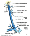
Internal Jugular Vein (IJV)

4 fun facts!
A
  1. Dilated continuation of sigmoid sinus
  2. Exits skull through jugular foramen
  3. Enclosed by the carotid sheath in neck
  4. Joins subclavian v.; Subclavian v.+ IJV = Brachiocephalic v.
18
Q

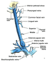
Name the 6 tributaries of the IJV:

A

– inferior petrosal sinus

– pharyngeal vv.

– occipital v.

– common facial v.

– lingual v.

– superior + middle thyroid vv.
19
Q

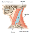
Blood Vessels in the Posterior Triangle?

(3)
A
  • Subclavian a. and branches
  • Subvlacian v.
  • EJV and tributaries
20
Q

Subclavian Artery in the Posterior Triangle:

  1. Where does the 3rd part of subclavian a. emerges from?
  2. What does it becomes after crossing lateral border of rib 1?
A
  1. between anterior and middle scalene mm.
  2. axillary a.
21
Q

Subclavian Artery in the Posterior Triangle:

  1. Where does the Transverse cervical a. branch from?
  2. What 3 muscles does it supply?
A
  1. branch of thyrocervical trunk (subclavian a.)
    * (passes laterally/posteriorly across base of posterior triangle, in front of anterior scalene m.)*
  2. supplies trapezius + rhomboids
22
Q

Subclavian Artery in the Posterior Triangle:

  1. Where does the Suprascapular a. branch from?
  2. What muscles does it supply?
A
  1. branch of thyrocervical trunk
    * (passes laterally across posterior triangle)*
  2. muscles on dorsal surface of scapula
23
Q

Subclavian Vein in the Posterior Triangle:

(5 fun facts)

  1. subclavian v. is a continuation of …?
  2. runs anterior to what structure in posterior triangle?
  3. passes anterior to what muscle?
  4. what major vein drains into the Subclavian v.?
  5. Subclavian v. + IJV = ???
A
  1. axillary v.
  2. subclavian a.
  3. anterior scalene m.
  4. EJV
  5. Brachiocephalic v.
24
Q

External Jugular Vein in the Posterior Triangle:

  1. what 2 veins converge to form the EJV?
  2. what muscle does the EJV cross before entering the posterior triangle?
  3. Where does it drain into?
A
  1. Posterior auricular v. + Retromandibular v. = EJV
  2. sternocleidomastoid m.
  3. subclavian v.

* NOTE: this is the ONLY thing you will find on the superficial surface of the SCM

25
Q

What are the anterior + posterior borders of the root of the neck?

What blood vessels are in the root of the neck?

A

Anterior = manubrium + upper margin of clavicle

Posterior = T1 + upper margin of scapula up to coracoid process

(Thoracic cavity projects into root of neck)

Blood vessels:

  • ● Subclavian a. and branches*
  • ● Subclavian v.*
26
Q

1st part of subclavian a.:

  • location
  • branches
A

LOCATION: ascends to the medial border of anterior scalene m.

3 main branches:

  • Vertebral a.
  • Internal thoracic a.
  • Thyrocervical trunk
27
Q

What are the 3 branches of the Thyrocervical trunk?

A

▪ inferior thyroid a.

▪ transverse cervical a.

▪ suprascapular a.

28
Q

2nd part of subclavian a.:

location?

A

passes laterally between anterior and middle scalene mm.

29
Q

3rd part of subclavian a.:

  • location?
  • becomes…?
A
  • crosses base of posterior triangle
  • becomes axillary a. after crossing rib 1 laterally
30
Q

Vertebral Artery

3 fun facts!

A
  • 1st branch of 1st part of subclavian a.
  • Goes through transverse foramina of C1-C6
  • Enters cranial cavity through foramen mangnum
31
Q

Internal Thoracic Artery

  • location?
  • what 2 branches does it give off?
A

LOCATION: Descends into thoracic cavity posterior to ribs and anterior to transversus thoracis m.

Gives off:

  • musculophrenic a. (to diaphragm)
  • anterior intercostal aa. (to thoracic wall)
32
Q

Thyrocervical Trunk: Inferior Thyroid Artery:

  • location?
  • innervates?
A

Ascends anterior and medial to anterior scalene m.

Supplies thyroid gland

*Superior continuation of thyrocervical trunk*

33
Q

Thyrocervical Trunk: Transverse Cervical Artery:

  • location?
  • branches?
A
  • Runs across anterior surface of anterior scalene m.

BRANCHES:

- Superficial branch continues on deep surface of trapezius m.

- Deep branch continues on deep surface of rhomboid mm.

34
Q

Thyrocervical Trunk: Suprascapular Artery:

  • location (runs across 4 structures)?
  • what fossa does it enter?
A
  • Runs across anterior surface of anterior scalene m. + phrenic n. + 3rd part of subclavian a. + trunks of brachial plexus
  • enters supraspinous fossa (after crossing over superior transverse scapular ligament)
35
Q

Subclavian Artery: Costocervical Trunk

The Costocervical trunk is just lateral to the thyrocervical trunk. What are its origins on the LEFT & RIGHT sides?

(hint: not the same)

A

Right: From 2nd part of subclavian a.

Left: From 1st part of subclavian a. (medial to anterior scalene m.)

36
Q

Subclavian Artery: Costocervical Trunk

  • location?
  • what are it’s 2 branches and where do they run to?
A
  • Runs posterior to anterior scalene m.

divides into two branches:

  1. Deep cervical a. ascends in the back of neck, and anastomoses with occipital a. (ECA)
  2. Supreme intercostal a. descends anterior to rib 1, and gives off posterior intercostal arteries 1 + 2
37
Q

Subclavian Artery: Dorsal Scapular Artery

  1. From what part of the Subclavian a.?
  2. what structures does it descend near?
A

1. 3rd segment

(could also be fr. thyrocervical trunk a.k.a. 1st segment)

2. along medial border of scapula deep to rhomboid mm

38
Q

Identify the branches of the common carotid artery:

A

1 = Common carotid a. (CCA)

2 = Internal carotid a. (ICA)

3 = Ascending pharyngeal a.

4 = Occipital a.

5 = Superficial temporal a. (Terminal branch ECA)

6 = Middle cerebral a. (branch of ICA)

7 = Anterior cerebral a.

8 = Middle meningeal a.

9 = Maxillary a.

10 = Facial a. (above lingual a.)

11 = Lingual a.

12 = External carotid a. (ECA)

13 = Superior thyroid a.

39
Q

CIRCLE OF WILLIS:

Identify the numbered branches

A
40
Q

Trace the blood flow paths to the circle of Willis for:

  1. the vertebral a.
  2. the internal carotid a.
A

1•Aorta → Aortic arch → Subclavian a. → Vertebral a. –> COW
2•Aorta → Aortic arch → Common carotid a. → Internal carotid a. –> COW

41
Q

Which artery in the Circle of Willis is particularily suseptable to strokes?

A

Middle Cerebral a.

42
Q

Subclavian Vein in the Root of the Neck

  1. is a continuation of what vein?
  2. what major vein drains into it (superiorly)?
  3. What vein joins the Subclavian v. to form the Brachiocephalic trunk?
A
  1. continuation of axillary v.
  2. External Jugular V.
  3. Joined by IJV (anterior to anterior scalene m) = Brachiocephalic v.
43
Q

External Jugular Vein (EJV): Tributaries

(diagram)

A
44
Q

Lymphatic Drainage of Head and Neck

match the nodes to their locations:

  • Deep cervical nodes
  • Pre-auricular and parotid nodes
  • Superficial cervical nodes
  • Mastoid nodes
  • Jugulo-digastric node
  • Jugulo-omohyoid node
  • Occipital nodes
  • Submental nodes
  • Submandibular nodes
A
45
Q

Superficial lymph nodes: Occipital

  1. drainage area?
  2. drains into?
A
  1. Posterior scalp and neck
  2. Superficial cervical nodes
46
Q

Superficial lymph nodes: Mastiod

  1. drainage area?
  2. drains into?
A
  1. Postero-lateral ½ of scalp
  2. Superficial cervical nodes

*receive lymph from retromandibular and pre-auricular nodes

47
Q

Superficial lymph nodes: Pre-auricular and Parotid

  1. drainage area?
  2. drains into?
A
  1. Anterior surface of auricle; Antero-lateral scalp; Upper ½ of face; Eyelids and cheeks
  2. Deep cervical nodes
48
Q

Superficial lymph nodes: Submandibular

  1. drainage area?
  2. drains into?
A
  1. Forehead + gingivae + teeth + tongue + structures along the path of facial a.
  2. Deep cervical nodes
49
Q

Superficial lymph nodes: Submental

  1. drainage area?
  2. drains into?
A
  1. Lower lip (central part); Chin and floor of mouth; Tip of tongue; Lower incisors
  2. Deep cervical nodes
50
Q

Superficial cervical lymph nodes drain into…

A

Deep cervical lymph nodes

  • forms chain around IJV
  • receives all lymphatic drainage from head + neck
  • (directly or via regional nodes)*
  • nodes form jugular trunks
51
Q

Jugulo-digastric node receives lymph from…

A

tonsils and tonsilar region

52
Q

Jugulo-omohyoid node receives lypmh from…

A

tongue

53
Q

Jugular trunks drain into…

A

THORACIC DUCT

  • forms in abdomen
  • enters thorax through aortic hiatus
  • terminates in the junction of left IJV and left subclavian v.
54
Q

THORACIC DUCT:

what 3 trunks join the jugular trunks here?

A

- left jugular trunk <– left side of head and neck

- left subclavian trunk <– left upper limb

- left bronchomediastinal trunk <– left ½ of thorax

55
Q

What 3 trunks drain into the junction of right IJV and right subclavian v.

A

- right jugular trunk <– right side of head and neck

- right subclavian trunk <– right upper limb

- right bronchomediastinal trunk <— right ½ of thorax + right upper intercostal spaces

56
Q

Cervical lymphadenopathy:

A
  • enlargement of cervical lymph nodes
  • manifestation of disease processes in head and neck
  • indication of diffuse diseases, e.g., lymphoma, sarcoidosis, glandular fever, HIV, etc.
57
Q

Node examination:

If there are soft, tender, inflamed nodes…

A

…Acute inflammatory process most likely to be infective

58
Q

Node examination:

If there are firm multinodular large rubbery nodes …

A

Lymphoma (mostly cancerous)