NECK - bones & fascia Flashcards

1
Q

Anterior & posterior boundaries of the neck?

A

anterior = lower border of mandible to upper surface of manubrium** **

posterior = superior nuchal line to intervertebral disc between C7 and T1 vertebrae

2
Q

inferior boundary of neck?

A

top of sternum to acromion

(encloses base of neck)

3
Q

Vertebral compartment contents?

(4)

A

Vertebral compartment:

Cervical vertebrae

muscles

spinal cord

cranial nerves

4
Q

visceral compartment contents?

(3)

A

Visceral compartment:

Larynx

pharynx

glands (thyroid, parathyroid, and thymus)

5
Q

Vascular compartment contents?

(2)

A

Vascular compartments (2 of them):

Major blood vessels

CN X (vagus n.)

6
Q

name the bony landmarks:

A
7
Q

which cervical vertebrae are considered “typical” ?

A

C3-C6

8
Q

C1 is also called?

it participates in what 2 major joints?

A

Atlas

atlanto-occipital joint (w. occipital condyles of cranium)

atlanto-axial joint (w. axis [C2])

9
Q

C2 is also called?

it participates in what major joint?

A

Axis

atlanto-axian joint (w. atlas [C1])

10
Q

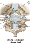
Name the components:

A
11
Q
  1. Name the 3 attachments of the cruciate ligament?

(superior, inferior, transverse)

  1. What is the purpose of the transverse ligimant?
A

Superior longitudinal band → basilar part of occipital bone

Inferior longitudinal band → dorsal surface of C2 body

Transverse ligament → small tubercle on the medial aspect of the superior articular facet of C1 (wraps around the dens of C2)

2. Transverse lig. KEEPS DENS FLEXED

12
Q
  1. name the attachments of the Anterior atlanto-axial ligament?
  2. what is it a continuation of?
A
  1. Lower edge of ventral arch of C1 ↔ Front of C2 body
  2. Cranial continuation of anterior longitudinal ligament
13
Q
  1. name the attachments of the Posterior atlanto-axial ligament?
  2. what differentiates it from the posterior longitudinal ligament?
A
  1. Lower edge of dorsal arch of C1 ↔ Upper edge of laminae of C2 (arch to arch)
  2. Distinct from posterior longitudinal ligament (goes fr. Vertebral body to vertebral body)
14
Q
  1. name the attachments of the Alar ligament?
  2. what is its main function?
A
  1. Medial tubercles of occipital condyle ↔ Dens of C2
  2. Resists excessive rotation of head on axial dens
15
Q

Name the superior, anterior, & posterior attachments of the hyoid bone:

A

superior = floor of oral cavity

inferior = larynx

posterior = pharynx

(all via ligaments)

16
Q
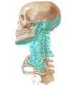
  1. Name the layers/sublayers of NECK FASCIA.
  2. name 3 functions of fascia.
A

Superficial fascia

Deep fascia

    • investing
    • pre-vertebral
    • pre-tracheal
    • carotid sheath

2. FUNCTIONS:

  • movements of neck (e.g. twisting)
    • swallowing
    • deep fascia controls spread of infection
17
Q

Name the fascia:

A
18
Q

Identify the fascial layers surrounding the 4 facial compartments:

A

1st compartment: area surrounded by investing layer

2nd compartment: area surrounded by pre-vertebral layer

3rd compartment (visceral compartment): area surrounded by pre-tracheal layer

4th compartment: area surrounded by carotid sheath

19
Q

the superficial fascia contains what muscle?

what is its action & innervation?

A

Platysma

muscle of facial expression (pulls the corners of mouth downward)

innervated by facial n. (CN VII)

20
Q

Neck: Investing Layer of Deep Fascia

name the 2 superior attachments:

A

external occipital protuberance

superior nuchal line
21
Q

Neck: Investing Layer of Deep Fascia

name the 4 inferior attachments:

A

scapular spine

acromion

clavicle

manubrium

22
Q

Neck: Investing Layer of Deep Fascia

name the 2 posterior attachments:

A

ligamentum nuchae

spinous process of C7 vertebra

23
Q

Neck: Investing Layer of Deep Fascia

name the 2 lateral attachments:

A

mastoid process

zygomatic arch

24
Q

Neck: Investing Layer of Deep Fascia

(facts)

A
  1. splits to enclose sternocleidomastoid and trapezius
  2. surrounds infrahyoid muscles anteriorly
  3. forms the roof of the posterior triangle of neck
  4. goes from base of skull to superior thorax
  5. forms fibrous capsule of parotid gland
  6. is pierced by blood vessels (external + anterior jugular vv.) and nerves (lesser occipital, great auricular, transverse cervical, supraclavicular nn., and all branches of cervical plexus)
25
Q

Neck: Pre-vertebral Layer of Deep Fascia

(facts)

A
  1. cylindrical layer surrounding vertebral column and associated muscles
  2. anteriorly attaches to anterior surfaces of transverse processes and bodies of C1-C7 vertebrae
  3. surrounds pre-vertebral muscles, scalene muscles (anterior, middle, and posterior), and deep back muscles
  4. axillary sheath surrounds brachial plexus and subclavian artery
26
Q

Neck: Pre-tracheal Layer of Deep Fascia

(facts)

A

● begins superiorly at hyoid and ends inferiorly in upper thoracic cavity

● individual components surround trachea, esophagus, and thyroid glands

● collectively known as “visceral fascia”

● buccopharyngeal fascia extends from base of skull to thoracic cavity

● buccopharyngeal fascia separates pharynx and esophagus from pre-vertebral layer

27
Q

Neck: Carotid Sheath

  1. receives contributions from what 3 fascial layers?
  2. name its 7 contents:
  3. infections can spread b/t what 2 locations?
A
  1. contribution from investing, pre-tracheal, and pre-vertebral layers

2. contents:

common and internal carotid aa.

internal jugular vein

vagus nerve

fibers from sympathetic trunk

carotid branch of CN IX

lymphatics

**3. between mediastinum & cranial cavity **

28
Q

Name the 3 fascial spaces and their locations:

A

1. Pre-tracheal space:

Between investing + pre-tracheal layers.

Passes between neck and anterior part of superior mediastinum.

2. Retropharyngeal space:

Between buccopharyngeal fascia + pre-vertebral fascia.

Posterior border is alar fascia.

Allows infection to spread from neck to mediastinum.

3. “Danger” space:

Between alar fascia + pre-vertebral fascia.

Extends from skull-base to mediastinum.

Conduit of infection between oropharynx/oral cavity and mediastinum/thorax.

29
Q

Neck Fasciae: Clinical Implications

(facts)

A
  • Prevention of spread of abscess by investing layer
  • Superior edge of manubrium is the lower limit for infections between investing layer and muscular part of pre-tracheal layer (surrounding infrahyoid muscles)
  • Spread into thorax of infection between investing layer and pre-tracheal layer (visceral part)
  • Abscess posterior to pre-vertebral layer can spread laterally into neck, causing a swelling posterior to sternocleidomastoid
  • Retropharyngeal abscess is caused by passage of abscess and pus from pre-vertebral layer to retropharyngeal space
  • “Danger” space is a passageway for infections between head/oropharynx/oral cavity and mediastinal structures (e.g., trachea, esophagus, etc.)
30
Q

Neck: Posterior Triangle

name the anterior & posterior borders; base, apex, floor, roof

A

● Anterior = Sternocleidomastoid

(posterior edge)

● Posterior = Trapezius

(anterior edge)

● Base = Clavicle (middle ⅓)

● Apex = Occipital bone posterior to mastoid process

● Floor = Pre-vertebral fascia

● Roof = Investing fascia

31
Q

Identify the muscles in the posterior triangle:

A
32
Q

what nerve(s) innervates the sternocleidomastiod mm., + trapezius mm.?

A

Accessory n.

(+ direct branches from C2-C3 ventral rami)

33
Q

what nerve(s) innervates the splenus capitus mm.?

A

Dorsal rami of cervical spinal nerves

34
Q

what nerve(s) innervates the levatator scapulae mm.?

A

Dorsal Scapular n.

35
Q

what nerve(s) innervates the anterior, middle, + posterior scalene mm.?

A

Adjacent ventral rami (C3-C7)

36
Q

what nerve(s) innervates the omohyoid mm.?

A

C1-C3 ventral rami

(via ansa cervicalis)

37
Q

Name the medial, lateral, + inferior borders of the anterior triangle:

A

Medial = midline of neck

Lateral = SCM (anterior edge)

Superior = inferior border of mandible

38
Q

What are the 4 sub-triangles located within the anterior triangle?

A
  1. Sub-mental Triangle (unpaired)
  2. Sub-mandibular Triangle (paired)
  3. Muscular Triangle* (paired)*
  4. Carotid Triangle (paired)
39
Q

Neck: Sub-mental Triangle:

  1. borders? (superior, inferior, lateral, floor)
  2. Contents? (2)
A

Borders:

Superior = mandibular symphysis

Inferior = hyoid

Lateral = digastric m. (anterior belly)

Floor = mylohyoid m.

Contents:

● Sub-mental lymph nodes

● Tributaries to anterior jugular v.

40
Q

Neck: Sub-mandibular Triangle

  1. borders? (superior, inferior)
  2. Contents? (5)
A

Borders:

● Superior = inferior border of mandible

● Inferior = digastric m. (anterior + posterior bellies)

Contents:

● Submandibular gland

● Submandibular lymph nodes

● Hypoglossal n. (CN XII)

● N. to mylohyoid

● Facial a. + v.

41
Q

Neck: Muscular Triangle

  1. borders? (superior, lateral, medial)
  2. Contents? (4 mm., 2 glands, 1 structure)
A

Borders:

● Superior = hyoid

● Lateral = omohyoid (superior belly) + SCM (anterior border)

● Medial = midline of neck

Contents:

▪ Sternohyoid + Omohyoid + Sternothyroid + Thyrohyoid

▪ Thyroid + parathyroid glands

▪ Pharynx

42
Q

Neck: Carotid Triangle:

  1. borders? (anterior, posterior, superior)
  2. Contents? (3aa., 2vv., 5CN, 2nn.)
A

Borders:

  • Antero-inferior = omohyoid (superior belly)
  • Superior = stylohyoid + digastric (posterior belly)
  • Posterior = SCM (anterior edge)

Contents:

▪ CCA + ECA and branches + ICA

▪ IJV + tributaries to common facial v.

▪ CN VII + IX + X + XI + XII

▪ Ansa cervicalis + Transverse cervical n.

43
Q

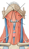
Name the suprahyoid mm. in the anterior triangle:

A
44
Q

What nerve(s) innervate the:

Digastric (posterior belly)

Stylohyoid

A

Facial n. (CN VII)

45
Q

What nerve(s) innervate the:

Digastric (anterior belly)

Mylohyoid

A

N. To mylohyoid

(branch of Inferior alveolar n. of CN V3)

46
Q

What nerve(s) innervate the:

Geniohyoid

A

Probably hypoglossal n. (CN XII)

47
Q

Name the infrahyoid mm. in the anterior triangle:

A

* from superficial –> deep:

1, 2, 3

48
Q

What nerve(s) innervate the:

Omohyoid (superior belly)

Omohyoid (inferior belly)

Sternohyoid

Sternothyroid

A

C1-C3 ventral rami (via ansa cervicalis)

49
Q

What nerve(s) innervate the:

Thyrohyoid

A

N. To thyrohyoid

[C1(2) ventral ramus(-i)]

50
Q

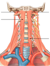
Name the Pre-vertebral and Lateral Vertebral Muscles:

A
51
Q

What nerve(s) innervate the:

Rectus capitis anterior

Rectus capitis lateralis

A

C1-C2 ventral rami

52
Q

What nerve(s) innervate the:

Longus colli

A

C2-C6 ventral rami

53
Q

What nerve(s) innervate the:

Longus capitis

A

C1-C3 ventral rami