Autolysis Flashcards

1
Q

Autolysis

A

自體溶解:self-digestion or degradation of cells and tissues by the hydrolytic enzymes(水解酶) normally present in those tissues

2
Q

Putrefaction

A

腐敗:postmortem bacterial metabolism and dissolution of host tissues (postmortem decomposition的細菌對宿主組織進行分解利用) 結果:color and texture changes, gas production, and odors

3
Q

Rigor mortis

A

屍僵: (1) 死後1-6小時肌肉產生強力收縮,通常持續1、2天直到用完所有ATP軟掉 (2) 若生物生前cachexia(惡病體質)、營養不良,則死後可能不會產生屍僵,因為沒有存夠足夠的ATP (3) 動物死亡後細胞內殘存的ATP與因全身性缺氧導致膜通透性改變,Ca2+大量流入細胞,繼續作用在肌肉(actin與myosin)上面使肌肉不正常收縮,而後因ATP的耗盡使myosin無法自actin上脫離,導致屍僵

4
Q

Algor mortis

A

屍冷:死後循環消失,導致體溫逐漸流失。可藉體溫判斷動物死亡多久 不能用在爬蟲類

5
Q

Livor mortis (hypostatic congestion)

A

屍斑:gravitational force –> 將血液往側躺方向拉產生紅色,而重量的壓迫產生白色,故會有紅白相間的屍斑,可幫助判斷死者的死亡姿勢,對法醫學第一現場的判斷很重要

6
Q

Chicken fat

A

Result of postmortem clotting血液凝集:

  1. 死後血液裡RBC與plasma成分紅白分明的凝集塊,一抽出來就斷裂,為其中一個區分柱塞的方法,又稱為chicken fat clot
  2. 正常健康的心臟,在死前一刻會急遽收縮擠出血液,使心臟內殘留的血液很少,不易產生chicken fat,假如死後有明顯的chicken fat產生,代表動物生前生理狀態差、身體虛弱,心臟無法強力的收縮,心臟內殘留的血液很多,因此出現在心臟的chicken fat clot對動物的死亡分析是有意義的。
  3. 反觀,出現在血管中的則是沒意義的,為正常死後變化
7
Q

hemoglobin imbibition

A

血紅素浸透:

  • pinkish to reddish coloration imparted to tissues due to the lysis of red blood cells.
  • beginning some hours after death
  • 不會用肉眼確認是否病變或死後變化,會在切片下確認
  • especially the heart and arteries (particularly evident in the aorta) and veins
8
Q

bile imhibition

A
  1. 動物死後幾個小時之內,膽汁會開始穿透膽囊並將周圍組織染黃,並逐漸轉為棕綠色
  2. 右肝藏呈棕綠色
9
Q

Pseudomelanosis

A

偽黑病變

致屍體呈現藍綠色的iron sulphide,因為:

(1) Fe in the Hemoglobin released by lysed erythrocytes reacts with
(2) H2S(硫化氫) generated by腐敗菌 (putrefactive bacteria) on the animal

* 不是因黑色素(melanin)所導致

10
Q

bloating

A

鼓脹

  1. 動物死後體內的細菌(尤其在GI tract)持續發酵產氣導致的結果,草食性動物相較於肉食性動物在死後鼓脹的較快也較嚴重
  2. 在死掉的反芻動物與馬的胃腸中,細菌會持續進行發酵,故死後不久因不會噯氣肚子鼓漲,為明顯的腹內postmortem decomposition,而在太陽照射下會加快bloating現象。
  3. 判斷是否為死後鼓脹,可由觀察食道或氣管上的鼓脹線來做為依據。

(i) 若為死前的鼓脹,屬於急性的變化,氣管或食道的後段會因腹壓擠壓而壓破血管,使血液受阻,呈現缺血後較白色的顏色,胸腔則因還有血液,黏膜會是正常因此呈現紅色

11
Q

Pale foci subserosally

A

漿膜下蒼白小點- 發生在肝臟

  1. 因腹腔內壓(intraabdominal pressure)上升,將血液擠壓出去
  2. bacterial action:細菌將組織降解,切片下沒有細胞核像凝固樣壞死(coagulation necrosis),沒有炎症反應且出現很多細菌,可判斷為死後變化
  3. 外觀:使肝臟表面上出現白色小點區域,變得不圓滑
12
Q

Mucosal sloughing

A

黏膜脫落Mucosal sloughing

保護腸道的黏液死後停止分泌,消化酵素開始分解腸道,使黏膜上皮細胞全面性脫落,屬死後自溶現象

13
Q

lens opacity

A

Lens opacity occurs when the carcass is very cold or frozen. The change will reverse to normal transparency on warming, but it can be confused with cataracts in cold carcasses.

水晶體混濁lens opacity:因缺氧導致蛋白質變性,變得混濁可藉由水晶體的混濁程度判斷該動物死亡多久,死亡愈久,水晶體愈混濁。易與cataracts白內障混淆,要判斷為何者仍須透過切片儀器判讀

14
Q

Atrophy and possible causes

A

萎縮

  • decrease in size, amount of cell or tissue or organ

•Disuse atophy

  • Overwork or persistent cell injury
  • Loss of innervation (denervation atrophy)
  • Loss of hormonal (trophic) stimulation
  • Reduced blood supply / hypoxia
  • Inadequate nutrition
  • Compression
  • Aging (senile atrophy)
  • Starvation and malnutrition atrophy
15
Q

What are the reversible cellular adaption?

A

Atophy, hyperplasia, hypertrophy, metaplasia

16
Q

Serous atrophy of fat

when happens?

A

脂肪細胞的萎縮

  • fat deposits are completely or partially depleted, and a clear or yellowish gelatinous material remains.
  • indicate starvation
17
Q

Involution

A

退化/生理性萎縮

  • normal physiological atrophy
  • eg Thymus, uterus after parturition
18
Q

Hypertrophy

especially for what types of cells?

A

肥大

  • for cells that undergo limited cellular proliferation/division, such as cardiac and skeletal muscle cells
  • no increase in number of cells, but increase in size and number of organelles
19
Q

Compensate hypertrophy

A

hypertrophy when

  1. loss of a part of an organ or one of the paired organs or
  2. from obstruction of the lumen of a hollow muscular organ.
20
Q

Hyperplasia

A
  • 增生: increase in number/layers of cells
  • 增生是排列整層增生,沒有影響外觀
  • Ability: Labile cells (epidermis, intestinal epithelium, and bone marrow cells)>Stable cells(bone, cartilage, and smooth muscle)> Permanent cells (neurons and cardiac and skeletal muscle myocytes)
21
Q

Pituitary Cushing Syndrome vs Adrenal Cushing Syndrome

A

Pituitary Cushing Syndrome

tumor in anterior pituitary gland–> over production of ACTH

–> which in turns over stimulates adrenal gland to produce cortisol

  • final result: adrenal gland 成個腎上腺cortex都變厚

Adrenal Cushing Syndrome

  • tumor in adrenal gland causing over production of cortisol
  • adrenal gland 變到好醜,亂長,難看到分界
22
Q

Metaplasia

Give one example

A

化生: one adult cell type is replaced by another adult cell type of the same germ line

Example: Smoking causes the psedostratified epithelium of airways to undergo metaplasia into stratified squamous epithelium under chronic irritation.

–> loss of cilia, less protective against antigen

Example2: lack of vitamin A –> transitional epithelium in urinary tract, epithelium of mucous gland of esophagus into squamous epithelium

23
Q

Dysplasia

A

異生dysplasia:腫瘤的前驅,細胞生長不固定方向且大小不一。在異生一開始時是有機會可逆的,此時basement membrane(基底膜)仍完整,若不改善則,一旦穿過basement membrane,終會走向腫瘤一途

24
Q

Storage disease

A

genetic defeats in transportation, protein folding etc resulting in lack of critical enzyme in breaking down and removing the substances that accumultaes in the lysosome

25
Q

Fatty change and the corresponding mechanisms in leading it

how is it in histology?

A

=hepatic lipidosis

Accumulation of triglycerides(三酸甘油), cholesterol (膽固醇)and phospholipids(磷脂) in the hepatocytes of liver

  1. Excessive delivery of free fatty acids either from the gut or from adipose tissue
  2. Decreased β-oxidation of fatty acids to ketones and other substances because of mitochondrial injury (toxins, hypoxia)
  3. Impaired synthesis of apoprotein (CCl4 toxicity, aflatoxicosis)
  4. Impaired combination of triglycerides and protein to form lipoprotein (uncommon)
  5. Impaired release (secretion) of lipoproteins from the hepatocyte (uncommon)

In histology, many haptocytes look like fat cells, and nucleus 會被擠到邊邊去,甚至看不到

26
Q

Fatty infiltration

A

Proliferation/hyperplasia of adipocytes, not fatty acid

muscle cell atrophy may result in infiltration of adipocytes

27
Q

Glycogen deposition

A

Excessive amounts of glycogen are present in animals in which glucose or glycogen metabolism is abnormal, such as diabetes mellitus, or in animals that have received excess amounts of corticosteroids.​

  • liver enlarged, 米黃色(beige to tan-white), non-greasy, do not float on formalin
    histology: vacuoles present (but irregularly in shape with indistinct outline) , nucleus centrally located, 蜘蛛網狀
28
Q

Proteinuria

A

蛋白尿

29
Q

Russel bodies

A
  • hyaline inclusion bodies present in plasma cell –> Matt cells
  • large, eosinophilic, homogenous, amorphorus
  • found in Spleen
30
Q

Protective unfolded protein response

A
31
Q

Crystalloid

A

Crystalloid: Crystalline protein 水晶狀蛋白質

  • intracellular inclusion bodies 核內包含體
  • 長條形在核中心
  • accumulation of 老化了的蛋白質
32
Q

The Splendore-Hoeppli Reaction

A
  • caused by persistant infection by foreign antigen,尤其是黴菌與特殊細菌
  • chronic(慢性)肉芽腫性inflammation response(炎症反應),產生許多抗體/補體和病原作用,形成immno complex(免疫複合體),稱Splendore-Hoeppli material
33
Q

hyaline microthrombi

A
  1. 玻璃樣微血管血栓hyaline microthrombi:DIC (disseminated intravascular coagulation) 血管內不正常的凝集,所有身體裡的微血管因為特殊原因(毒素、細菌的內毒素和燒傷等)而全面病變,全身微血管發生血液凝集時,此生物便會馬上缺氧休克而死

腫脹的包氏囊,肉眼看不出,需切片染色,會造成動物急性death

34
Q

hyaline membranes of the alveolar

A
  1. 肺泡的玻璃樣膜hyaline membranes of the alveolar:專指肺泡微血管因病毒感染造成cytokine釋放造成肺炎,導致通透性過度增加,使蛋白質大量流出在肺泡表面堆積,造成呼吸困難,生物體最後因呼吸衰竭而死
35
Q

Amyloid

A

類澱粉質

  1. 成分為摺疊不完全的蛋白質(misfolded proteins),因蛋白質上有醣基化修飾,滴入iodine(碘液)會呈現黑紫色如澱粉而得其名
  2. 會沉積在微血管末梢,如peripheral arteries(周末動脈)、glomerulus(絲球體)和sinuloid(竇狀隙),容易導致周遭血管物理性的堵塞或壓迫,導致atrophy、缺血進而壞死,堆積在絲球體的臨床症狀為蛋白尿proteinear
  3. 最常使用的特殊染色為剛果紅Congo Red,在偏光鏡polarized light帶有青蘋果螢光綠
  4. Amyloid產生其因細胞內β-sheet蛋白質不正常的產生,兩兩相遇後會polymerase越拉越長,形成長形絲狀物,與剛果紅特殊染色(可與DIC做區別)有關
36
Q

Secondary Amyloidosis

A
  • under chronic inflammation,
  • 是動物最普遍的患病形式
  • 這些amyloid可能會沈積在kidney/liver/spleen/lymph node
  • 最重要為沈積在腎臟Kidney,因為過不到glomerulus(絲球體),一直沈積,造成物理性堆積,導致缺氧hypoxia,內皮細胞受損,蛋白質流出,導致動物的proteinuria(蛋白尿)
37
Q

vasculitis

A

血管炎

38
Q

Beta-amyloidosis

A

在人的病例中,胞外堆積amyloid-beta protein (Aβ)導致了Alzheimer’s disease(阿茲海默症);老狗腦中亦發現此堆積,有Aβ plaques,但在其中動物身上比較少見

39
Q

fibrinoid change

A

類纖維蛋白變性

  • result of the deposition of immunoglobulin, complement, and/or plasma proteins, including fibrin in the wall of a vessel.
  • This lesion is due to injury to the tunica intima and media such as occurs in the immune-mediated vasculitides血管炎.
40
Q

Gout

A

通風

  • deposition of sodium urate crystals (尿酸鹽結晶)or urates in tissue.
  • It occurs in human beings, birds (kidney mostly), and reptiles but has not been reported in domestic animals.
  • Grossly, the serosa is covered with a thin layer of gray granules關節腫漲或臟器表面呈白色粉粉的樣子
  • Microscopically, urate crystals have been dissolved during the preparation of paraffin- embedded histological sections, are visibly surrounded by numerous neutrophils, macrophages, and giant cells. (original 呈嗜伊紅性菊花狀結晶)
  • Physically harm –> 沉積在關節/臟器表面釋出的現象引發疼痛,
41
Q

Cholesterol crystals

A
  • caused by chronic inflammation/by product of hemorrhage and necrosis
  • resemble shards of glass
  • 屬於油類物質,在製作切片酒精脫蠟時會被溶掉,外觀為裂隙狀
42
Q

dystrophic calcification

A
  • occurs in areas of necrosis-dead and dying cells can no longer regulate the influx of calcium into their cytosol, and calcium accumulates in the mitochondria. Common sites include necrotic myocardium, necrotic skeletal muscle, granulomas such as tuberculoid granulomas in cattle, and dead parasites,
  • Microscopically, calcium salts stain blue with hematoxylin and appear as fine amorphous granules or clumps, which can be either intracellular or extracellular. It is revealed more dramatically by special stains, such as von Kossa and Alizarin red S.
43
Q

metastatic calcification

A

blood calcium –> accumulaiton of Ca into cells, esp mitochondria

  1. 腎衰竭renal failure:腎衰竭會導致體內的磷(phosphates)滯留(體內鈣與磷的比例約為1:1),血磷高導致身體會想提升血鈣以維持比例,引發二次性副甲狀腺亢進(secondary renal hyperparathyroidism),副甲狀腺腫漲,分泌升鈣素(ex PTH)à血鈣過多(hypercalcemia)沉積於身體各處(gastric mucosa, kidney, alveolar septa),而骨骼中的鈣被提出造成骨質疏鬆
  2. Vitamin D toxicosis(維他命D中毒):草食獸食入促進鈣吸收的植物(如夜丁香),或是犬貓食入含有cholecalciferol的滅鼠藥,具有升鈣素的功能,或非夜行性動物在飼料中添加過多Vitamin D,導致過量血鈣增加
  3. 副甲狀腺素parathormone (PTH)和PTH-related protein:先天性副甲狀腺亢進(primary hyperparathyroidism)的病例較為稀少。犬隻罹患此病與惡性淋巴腺瘤(canine malignant lymphomas)和圍肛腺(canine adenocarcinoma of the apocrine glands of anal sac)有關(圍肛腺腫瘤會分泌PTH-related protein,具有提高血鈣的功能)
  4. 原發或轉移性腫瘤(metastatic neoplasms) 造成骨質的破壞,骨骼中的鈣跑到身體其他地方
44
Q

異位骨heterotopic/ectopic bone

A
  1. :異位骨化:骨頭形成在不正常的位置,可能顯示該地方曾經有發炎,分為兩類:
    1. 異位化生heterotopic metaplasia:骨細胞及組織出現在不對的位置,被認為起源於胚胎細胞的殘存,
    2. 骨質化生Osseous metaplasia:從不同種類的結締組織分化成骨母細胞osteoblasts形成骨質osteoid,其鈣化的形式與一般骨骼相同
45
Q

carotenoid pigments/lipochrome pigments

A

類胡蘿蔔色素

  • 草食動物脂肪裡沉積的天然色素,造成黃色脂肪, eg horses
  • 與黃疸icterus不同,此色素沉積是健康且正常的,區別為切片下不會有任何炎症細胞出現 (馬的脂肪因為吃草會比較黃,所以再去檢查mucosa,也要mucosal membrane黃才會是黃疸)
46
Q

tetracycline

A
  1. 四環素tetracycline:為抗生素,含有黃色色素;小動物在幼年時期若使用太多抗生素,在齒質生成時會將之染上黃色素,所以牙齒會顯示黃黃
47
Q

Lipofuscin-ceroid

A

脂褐素-蠟樣脂質

  1. Lipofuscin,又稱wear-and-tear pigment是巨噬細胞自噬反應後不能再被分解的最終產物。因之不能被溶體分解與行胞吐,所以會堆積在溶體中,是生物體廢物的一種形式
  2. Ceroid,與lipofuscin有許多相似的組織化學性質,但不同的是ceroid是_病理性大範圍_的組織發生自噬反應,產生大量lipofuscin的沉積,在胞外和胞內皆可能發生。常見在嚴重營養不良、缺乏維生素E與硒、惡病質、放射性治療等等情況中,被視為對細胞有不良的影響
48
Q

Edema

A

excess water runs into tissue from intracellular space or blood vessels

49
Q

hyperemia

A

Hyperemia充血

血管中血液過量增加,使血管較擴張但通透性正常。

eg demand for O2 changes over time

50
Q

congestion

A

鬱血

due to vein 被塞住了:通常指靜脈問題。為靜脈血回流受阻而淤積,導致hydrostatic pressure靜水壓上升。

51
Q

hemostasis

A

凝血

為密封受傷血管阻止流血的生理反應。

與Endothelium內皮完整性、Platelets、Coagulation cascade(凝血串聯反應)有關。

52
Q

hemorrhage

A

blood out of blood vessels and run into tissue

53
Q

Thromboisis

A

血栓

血管中形成的血塊blood clot,於循環系統中會妨礙或阻斷血流,通常為不正常活化凝血反應導致。

血栓若_剝落流動到更遠可能變成栓塞(Embolism)_ ex.貓若有血栓症候群可能就會突然地導致後肢癱瘓

54
Q

disseminated intraviscular thrombosis

A

瀰漫性血管內凝血

細菌毒素或全身性休克反應可能會導致體內微血管包括血竇(sinusoid)、肺臟、腎絲球體這些微小血管的不正常凝血,且通常是全面性地發生。Ischemia–>necrosis–>organ damage!

P.S. 並不是大條血管凝血才會嚴重!因人體大部分血液都在微血管,所以當微血管全面地發生凝血或出血的話會造成嚴重循環障礙

may run out of platelet and coagulation factor too, so unable to 凝血at other bleeding site

55
Q

Embolism

A

栓塞

血液中出現的異常物質阻塞血流

56
Q

Embolus

A

可阻塞血流之隨血液循環的不正常物質。

abnormal detached traveling intravascular mass, which is capable of blocking flow

57
Q

Infarction

A
  • hypoperfusion –> ischemia –> infarction
  • 血流減少引發的壞死區域。
  • 通常血拴(thrombosis)或拴塞(Embolism)導致血流阻塞,使大區域組織壞死造成梗塞(但若臟器有多套血管供應就較不會發生梗塞)
  • P.S.壞疽也是一種梗塞,其血液支配區域在末梢,發生大區域壞死
  • An area of necrosis secondary to decreased blood flow
58
Q

Shock

A

心血管衰竭或循環系統失靈。

血液循環系統中讓血液流動的血壓失去作用,使所有血液跑到全身微血管而回不了肺臟做交換,會導致休克

Cardiovascular collapse/failure of circulation

59
Q

Portal hypertension

A

Portal hypertension肝門靜脈高壓:

  1. 右心衰竭(heart failure)使血液積留於右心,血液淤積於靜脈;
  2. 肝硬化(hepatic cirhosis)導致腹水(ascites)或纖維化(fibrosis)綁住心血管系統,使血液回流受阻,導致靜水壓上升
60
Q

Exudate, Transudate

A
61
Q

hepatic cirrhosis

A
  • consistent inflammation in the liver, leading to accumulation of scar tissues
  • fibrosis too severe–>cirrhosis
62
Q

ascites

A

腹水/hydroperitoneum

  1. 較易由外觀判斷(如下圖)。
  2. 通常胸水會讓動物喘得較嚴重,但腹水也可能會因腹壓過大影響橫膈運動而讓動物喘。
  3. X光可見比較難穿透,用超音波可更容易看出水分成分
63
Q

hydrothorax

A

胸水

  1. 較難由外觀判斷,要從X光穿透力變差去判斷可能有積水。
  2. 動物較容易喘。
64
Q

Hydropericardium

A

心包囊積液

65
Q

Effusions

A

積液

  • Increased fluid in the body cavities
  • 可用於鳥類、烏龜等沒有胸、腹只有體腔的動物。
  • 可能為Transudate或Exudate
66
Q

Anasarca

A

全身水腫

  • 循環障礙
67
Q

Pitting Edema

A

Pitting Edema凹陷性水腫

觀察外觀疏鬆組織,疑似可能是水腫時可用按壓去檢測是否真正是水腫。若此處只是胖,按壓脂肪並不會留下指痕。

  1. 正常組織因有彈性所以會很快恢復,不會留下凹陷壓痕。
  2. 水腫的部位需要相對久的時間回復,因為組織中彈性纖維所在處  被水分塞滿了,彈性較差。
68
Q

Brisket Disease

A
  • 反芻獸很好觀察的一種水腫臨床徵狀

反芻獸因進食時瘤胃本來就很大充滿液體,所以要用最靠近地心引力的地方判斷,通常水腫液體會累積在脖子、胸前這塊的結締組織中。

69
Q

What does it look like in histology of pneumonary edema?

A
  1. 怎麼顯微鏡下肺泡呈現一片嗜伊紅性蛋白?(圖三下方)

A: 顯微鏡下肺泡腔內可看到很多粉紅均質物質,因為肺泡內會有type Ⅱ pneumocytes分泌的表面張力素等(為蛋白質樣物質)去和液體混和,如此反而方便我們在顯微鏡下的判讀。

70
Q

hypoalbuminemia

A

低蛋白血症

肝硬化的肝細胞失去正常功能,無法合成白蛋白albumin

   –> 血液中白蛋白減少導致低蛋白血症

–>進而降低血管內膠體滲透壓而水腫

71
Q

Traumatic Reticulopericarditis

A

創傷性的蜂巢胃心外膜炎 –> Brisket Edema

72
Q

Pulmonary hypertension syndrome in broiler chickens

A
73
Q

subcutis edema

A

皮下水腫

74
Q

Cyanosis

A

發紺:Cyanosis is the bluish or purplish discolouration of the skin or mucous membranes due to the tissues near the skin surface having low oxygen saturation.

75
Q

arryhythmia

A

心律失常

76
Q

nutmeg liver

A

荳蔻肝

77
Q

siderotic plaque

A

脾臟鐵末班塊

肉眼見脾臟上有黃褐色硬實物質(Brown induration),即巨噬細胞吞老舊紅血球所形成的Hemosiderin-laden macrophages。

此為動物鬱血證據,可推測發生局部性脾臟扭轉導致的血液淤積,或是全身性鬱血(可懷疑心衰竭)

78
Q

Hemophilia

A

Hemophilia血友病: 容易出血。人較常出現,而動物常出現的為狗豬馬。

  1. Hemophilia A-factor VIII:缺乏factor VIII。發生於狗馬豬
  2. Hemophilia B-factor IX:缺乏factor IX。發生於狗
79
Q

hematoma

A
80
Q

Dissection

A

剝離

81
Q

Petechiae

A
82
Q

Purpura

A

紫瘢

  • <1 cm的出血點。
  • 可見心臟冠狀溝有很多出血點,跟中毒有關
  • purple-colored spots that are most recognizable on the skin. The spots may also appear on organs or mucous membranes, including the membranes on the inside of the mouth.
  • Purpura occurs when small blood vessels burst, causing blood to pool under the skin. This can create purple spots on the skin that range in size from small dots to large patches.
83
Q

Ecchymoses

A

斑狀出血: >1 cm 的出血點( subcutaneous spot of bleeding),即Bruise皮下瘀傷(瘀青)

the same as purpura: purpura is <1cm

Specifically, bruises(hematoma) are caused by trauma whereas ecchymoses, which are the same as the spots of purpura except larger, are not necessarily caused by trauma,[3] often being caused by pathophysiologic cell function, and some diseases such as Marburg virus disease.

84
Q

Thrombocytopenia

A

血小板減少

85
Q

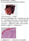
Cardiac Tamponade

A

心包填塞

圖為切開心包膜後裸露的心臟。切開後發現充滿大量血液。可能是動脈或血管發生破裂,即跟血管病變有關。血液大量在心包囊(pericardium)聚集,導致心包填塞(tamponade),心臟無空間可以跳動,會使全身突然地灌流量不足而導致休克,動物會突然死亡。

86
Q
A
87
Q

coagulative necrosis

A

The microscopic anatomy shows a lighter staining tissue (when stained with H&E) containing no nuclei with very little structural damage, giving the appearance often quoted as ‘ghost cells’. The decreased staining is due to digested nuclei which no longer show up as dark purple when stained with hematoxylin, and denaturation of intracellular proteins will somehow give them a darker pinkish stain eosin.

88
Q

infarct

A

梗塞

89
Q

Coagulative necrosis

A

凝固氧壞死

  1. maintenance of normal architecture of necrotic tissue for several days after cell death.
  2. the default pattern of necrosis associated with ischemia or hypoxia in every organ in the body except the brain.
  • Gross Appearance: tissue is firm and architecture is maintained for days after cell death.
  • Microscopic: Preserved cell outlines without nuclei.
90
Q

Liquefactive necrosis

A

液化壞死

  1. The pattern of necrosis seen with infections.
  2. Also, the pattern is seen following ischemic injury in the brain
  3. The release of digestive enzymes and constituents of neutrophils is the reason for liquefaction in infections.
  • Gross Appearance: The tissue is in a liquid form and sometimes creamy yellow because of pus formation.
  • Microscopic: Inflammatory cells with numerous neutrophils.
91
Q

Fat necrosis

A
  • acute inflammation affecting area of numerous adipocytes, eg pancreas, breast,

-

  1. 和胰臟有關,會有此引發急性胰臟炎(犬貓特別多)
  2. 胰液滲出後周圍細胞被胰液中的脂肪酶分解,形成腹痛、腹部肌肉僵硬、嘔吐
  3. 雖然胰臟炎發生在腹下,但滲出的lipase會跟著血液擴散到全身,影響到皮下脂肪,造成皮下脂肪炎(皮下會硬硬的)
92
Q

lipofuscin

A
  • wear and tear pigment
  • fine yellow-brown pigment granules in cytosol
  • composed of lipid-containing residues of lysosomal digestion, eg after autolysis

-

93
Q

Jaunice

A

= Icterus黃疸

  • imbalance bewteen formation and removal of bilirubin
  • Mucous membrane(眼白,虹膜等等)呈黃色,結締組織或脂肪組織都被染成黃色。肉眼可見,切片除肝臟外見不到
  1. 肝前性 (Prehepatic) :過多的 bilirubin 的產生、紅血球破壞過多、肝功能正常,通常與溶血性疾病有關。像血液內寄生蟲,病毒性感染貧血,大範圍脾臟出血也會有肝前性黃疸
  2. 肝因性 (Hepatic) :肝細胞受損減少肝細胞功能,白蛋白合成受損,代謝 bilirubin-albumin complex 的功能不足。 判斷:看看肝臟有沒有壞死或是先天性問題,臨床上肝硬化或肝腫瘤都會出現黃疸
  3. 肝後性 (Posthepatic) :膽管系統的阻塞使膽汁無法排入小腸。Ex.膽管內的肝吸蟲、膽管炎、膽囊發炎都會產生
94
Q

Porphyria

A

紫質

造成疾病的主因是血基質(heme) 的前驅物-紫質(porphyrin)及其衍生物代謝異常,使患者體內的紫質或其前驅物累積過量而致病。

95
Q

Gastric dilation volvulus (GDV)

A
  1. Torsion內臟扭轉:

通常大動物吃很飽後激烈運動(多為姿勢的劇烈變化,如警犬剛吃飽趴在地上立刻接受指令坐起來)立刻會導致內臟翻轉,使血液在此處

淤積回不到心臟。不但使內臟疼痛腫大,可以回右心再往體循環的血量也變少,動物很快會因低血量休克(hypovolemic shock)死亡。

–> acute local congestion

96
Q

Brown induration或Hemosiderin plaque

A
97
Q

how the followings cause hemostasis disorder?

  1. Anthrax
  2. cirrhosis
  3. thromboopenia
A
  1. 感染Anthrax炭疽導致敗血症,進而anoxemia(低血氧)而血液無法正常凝集。
  2. Prothrombin, fibrinogen, factor VII, and factor X數量下降導致凝血不全。所以肝硬化病人容易出血。
  3. insufficient platelets, causing hemophilia